

Subscribe to RSS
DOI: 10.1055/s-0041-1730956
Synthesis and Antitumor Activity of (3-Hydroxyacrylato-O,O′) Diammineplatinum(II)
Authors
Funding This work was supported by the Shanghai Innovation Action Plan of Science and Technology (Grant No. 14431905900). We thank Dr. MA Jing for support.

Abstract
As an indispensable part of cancer chemotherapy, platinum drugs still play an important role in cancer treatment. In this study, two platinum(II) complexes with Michael acceptor 3-hydroxyacrylic acid as the leaving group were synthesized from cis-diamminediiodo platinum(II) and 3-ethoxyacrylic acid. The structures of complexes 1 and 2 were confirmed by elemental analysis, infrared, 1H NMR, 13C NMR, and HRMS (high-resolution mass spectrometry). Results from MTT assay showed that complexes 1 and 2 significantly inhibited the growth of the four human tumor cell lines (HCT-116, A549, CFPAC-1, and BxPC-3) with the IC50 values of the two compounds similar to that of the control drug (oxaliplatin) on HCT-116 and A549. Besides, results from an in vivo study in a mouse S180 sarcoma model showed that complex 1 had a higher antitumor activity in comparison to oxaliplatin. In conclusion, our article indicated that complex 1 deserved further research and development in cancer treatment.
Introduction
Cisplatin, a platinum(II) complex, has made a major impact in the chemotherapeutic treatment of testicular and ovarian cancers since the accidental discovery of its biological activity, and is widely used in the treatment of these types of cancers.[1] [2] However, it still has nonnegligible toxic and side effects such as nephrotoxicity, emetogenicity, and drug resistance, which cripple its overall effectiveness in cancer therapy.[3] [4] [5] [6] For decades, thousands of platinum(II) complexes have been prepared in the hope of finding those with more tolerable toxicological profile and higher efficacy.[7] These efforts have brought several new drugs (carboplatin, oxaliplatin, nedaplatin, and lobaplatin) into market,[8] [9] [10] followed by several new complexes emerging in current clinical trials ([Fig. 1]).[11]
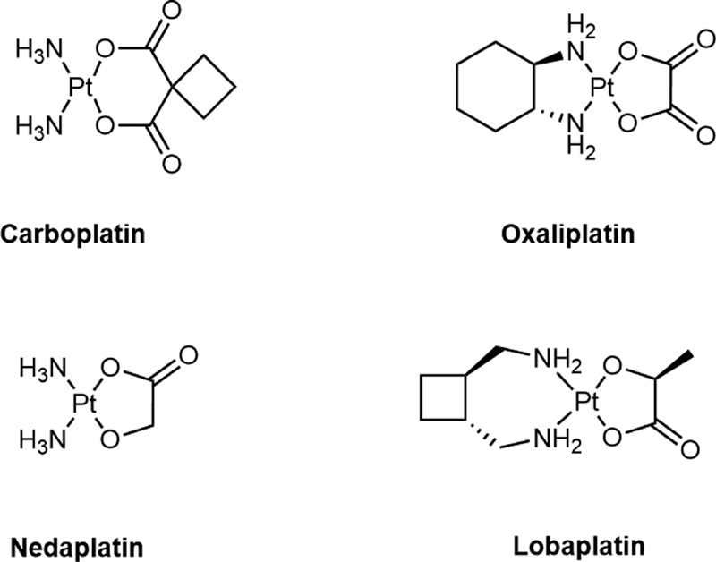
With regard to most of the platinum(II) complexes, such as carboplatin and oxaliplatin, dicarboxylate plays a role as the leaving group in the mechanism of the interaction between platinum(II) complex and DNA, and in addition to platinum(II) complexes containing dicarboxylate as the leaving group, there are also platinum(II) complexes, such as nedaplatin and lobaplatin, containing α-hydroxylcarboxylate as the leaving group, which will have more stronger antitumor activity.[12] Thus, exploring a novel compound based on the structure of platinum(II) complexes containing α-hydroxylcarboxylate may represent an promising strategy to improve the antitumor activity of the platinum(II) complexes.[12]
To the best of our knowledge, the synthesis of platinum(II) complexes with Michael acceptor as leaving groups has not been reported. Michael acceptor is the functional group in which the olefins or acetylenes conjugated to electron-withdrawing groups. The compounds with Michael acceptor are considered as a class of biologically active molecules which directly or indirectly involved in the life processes. A series of studies suggested that the Michael acceptor moiety especially α,β-unsaturated carbonyl fragment is the essential active group with cytotoxicity among various anticancer compounds.[13] [14] [15] [16] Michael acceptor is a fragment for covalent binding and might improve the nonselective cytotoxicity of platinum(II) complexes. In this study, two 3-hydroxyacrylatoplatinum(II) complexes containing Michael acceptors as leaving groups (complexes 1 and 2) were synthesized and characterized ([Fig. 2]). Our data suggested the potential use of these two compounds in cancer treatment in the future.

Results and Discussion
Successful Synthesis of Complexes 1 and 2
[Scheme 1] shows the synthesis of complexes 1 and 2 following a general method.[17] Starting from cis-[PtR2I2] (commercially available), the first step was performed in water with AgNO3 to form [PtR2(H2O)2](NO3)2. Furthermore, [PtR2(H2O)2](NO3)2 was mixed with sodium 3-ethoxyacrylate to produce a yellow solution. The reaction mixture was concentrated in vacuum and purified by silica gel chromatography to obtain the target complex. The products were then characterized by elemental analysis, infrared (IR), 1H NMR, 13C NMR, mass spectrometry, and high-resolution mass spectrometry, respectively. The elemental analysis data for each compound were in good agreement with the designed structure formula. The binding of the 3-hydroxyacrylate to platinum atoms as a bidentate ligand was confirmed by the shift of νC=O to lower frequencies and the absence of νO-H absorption in IR spectra in the resulting complexes.[4] All complexes showed [M + H]+ peaks, corresponding to their molecular weights, and had three typical protonated molecular ion peaks reflecting the platinum isotopes: 194Pt(33%),195Pt (34%), and 196Pt (25%). 1H and 13C NMR spectral peaks matched the chemical structures given in [Fig. 2]. At room temperature, the solubility values of complexes 1 and 2 in phosphate buffered saline (pH = 7.4) are 36.5 and 1.5 mg/mL, respectively. Complex 1 possesses sufficient water solubility.

An In Vitro Study Showing Cytotoxic Activities of Complexes 1 and 2 on Cancer Cells
In this study, the in vitro cytotoxicity of the platinum(II) complexes was tested by measuring the effect of the complexes on the proliferation of the four cancer cells (the colorectal cancer cell line HCT-116, the lung carcinoma cell line A549, as well as the pancreatic cancer cell line CFPAC-1 and BxPC-3). These four cell lines were continuously exposed to different concentrations of oxaliplatin (serving as a control drug), as well as complexes 1 and 2 for 48 hours, and then IC50 of the three drugs was assessed using MTT assay according to a reported study.[18] [Table 1] shows that both complexes 1 and 2 showed cytotoxic activity on the four cell lines with the significant effect being seen in HCT-116 and A549 cells. Interestingly, the cytotoxicity of complex 1 was less and closer to that of the control drug when compared with complex 2. Thus, we chose complex 1 for the following study.
Complex 1 Inhibited Tumor Growth in a Mouse S180 Sarcoma Model
The antitumor activities of complex 1 and oxaliplatin were further compared in a mouse S180 sarcoma model.[19] Based on preliminary studies, tumor-bearing mice were administered with intraperitoneal injection of complex 1 (25 mg/kg) once-daily. [Table 2] shows that complex 1 displayed a strong antitumor effect (tumor growth inhibition: 69.78%). However, administration of complex 1 also led to severe mouse weight loss, suggesting that the mice could not tolerate once-a-day administration of complex 1, and the safe and effective dosage regimen should be explored.
|
Group |
Dose (mg/kg) |
Dosing regimen |
Mean body weight (g) |
Tumor weight (g) |
TGI (%) |
|
|---|---|---|---|---|---|---|
|
D1 |
D7 |
|||||
|
Control |
Vehicle |
once-daily |
18.88 ± 0.97 |
21.45 ± 1.83 |
3.21 ± 0.33 |
/ |
|
Complex 1 |
25 |
once-daily |
19.21 ± 0.94 |
15.44 ± 0.90 |
0.97 ± 0.35 |
69.78** |
Abbreviation: TGI, tumor growth inhibition.
a Tumor-bearing mice were treated by intraperitoneal (ip) injection of complex 1 for 7 days. Data are presented as mean ± SD. The comparison between the two groups was conducted using t-test with statistically significant at **p < 0.01 versus control.
Increased Drug Given Dose and Extended Drug Given Interval May Enhance Antitumor Effect While Reducing Toxicity of Complex 1 in Mouse Xenograft Models
Based on the results obtained from [Fig. 2], we further assessed whether decreasing complex 1 dose (15 mg/kg, once-daily) or prolonging its dosing interval (30 mg/kg, once in 2 days) will improve the effect of complex 1 on mouse body weight and tumor weight. [Table 3] shows the antitumor effect of complex 1 when administered at 30 mg/kg; once in 2 days was more effective than daily dose at 15 mg/kg, while the mouse weight loss of the complex was less severe. Interestingly, although the antitumor effect of complex 1 (30 mg/kg, once in 2 days) was weaker than the control drug (oxaliplatin, 9 mg/kg) at a same dose interval (once in 2 days), the toxicity of compound 1 was significantly reduced.
|
Group |
Dose (mg/kg) |
Dosing regimen |
Mean body weight (g) |
Tumor weight (g) |
TGI (%) |
|
|---|---|---|---|---|---|---|
|
D1 |
D11 |
|||||
|
Control |
Vehicle |
Once-daily |
21.18 ± 0.91 |
24.08 ± 3.32 |
2.95 ± 0.38 |
/ |
|
Complex 1 |
15 |
Once-daily |
20.94 ± 0.64 |
18.82 ± 1.08 |
1.66 ± 0.39 |
43.58** |
|
Complex 1 |
30 |
Once in 2 days |
20.86 ± 1.13 |
20.21 ± 3.22 |
1.60 ± 0.35 |
45.87** |
|
Oxaliplatin |
9 |
Once in 2 days |
21.14 ± 1.04 |
17.46 ± 1.85 |
1.18 ± 0.24 |
59.85** |
Abbreviation: TGI, tumor growth inhibition.
a Tumor-bearing mice were treated by intraperitoneal (ip) injection of complex 1 for 11 days. Data are presented as mean ± SD. The comparison between the two groups was conducted using t-test with statistically significant at **p < 0.01 versus control.
Then, we further increased drug dose and extended drug dosing interval, and investigated whether a more effective and safer use of compound 1 would be achieved when compared with the control drug (oxaliplatin, 9 mg/kg, once in 2 days). Thus, the drug dosing interval was increased to once in 3 days (60 mg/kg) or once in 6 days (120 mg/kg) in the mouse xenograft models. As shown in [Table 4], bolus application of compound 1, both 60 mg/kg, once in 3 days, and 120 mg/kg, once in 6 days, exhibited stronger antitumor activity than oxaliplatin with mouse weight being preserved even on 11th day, suggesting better safety profile of a larger dose at a longer interval of complex 1 at the two dosage regimens. Interestingly, a single bolus of complex 1 at 120 mg/kg once in 6 days gave the best result.
|
Group |
Dose (mg/kg) |
Dosing regimen |
Mean body weight (g) |
Tumor weight (g) |
TGI (%) |
|
|---|---|---|---|---|---|---|
|
D1 |
D11 |
|||||
|
Control |
Vehicle |
Once in 3 days |
22.81 ± 1.17 |
24.56 ± 3.67 |
3.08 ± 0.32 |
/ |
|
Complex 1 |
60 |
Once in 3 days |
22.21 ± 0.73 |
22.46 ± 2.28 |
1.10 ± 0.56 |
64.39** |
|
Complex 1 |
120 |
Once in 6 days |
22.78 ± 0.75 |
20.34 ± 2.44 |
1.02 ± 0.36 |
66.83** |
|
Oxaliplatin |
9 |
Once in 2 days |
23.18 ± 0.62 |
19.92 ± 2.50 |
1.16 ± 0.20 |
62.42** |
Abbreviation: TGI, tumor growth inhibition.
a Tumor-bearing mice were treated by intraperitoneal (ip) injection of complex 1 for 11 days. Data are presented as mean ± SD. The comparison between the two groups was conducted using t-test with statistically significant at **p < 0.01 versus control.
Conclusion
In summary, two 3-hydroxyacrylatoplatinum(II) complexes with novel six-membered ring structures containing Michael acceptors as leaving groups were synthesized and characterized. Both complexes 1 and 2 were evaluated for cytotoxicity against four human cancer cell lines and complex 1 was evaluated for antitumor activity in a mouse S180 xenograft model. The results showed that the anticancer effects of complexes 1 and 2 were similar to that of oxaliplatin in two human cancer cell lines. Furthermore, we explored the different dosing regimens of complex 1 in an in vivo study. Our data showed that administration of complex 1 at 120 mg/kg once in 6 days was more efficacious and safer than the control drug (oxaliplatin). In conclusion, 3-hydroxyacrylatoplatinum(II) complexes with novel six-membered ring structures containing Michael acceptors (complexes 1 and 2) at a higher dose and a longer interval may serve as promising drug candidates in cancer therapy.
Experimental Section
(3-Hydroxyacrylato-O,O′) diammineplatinum(II) (complex 1): To a solution of 3-ethoxylacrylic acid (1.0 g) in water (100 mL) was added NaOH (340 mg). The solution was then shaken ultrasonically, adjusted to pH = 7 by NaOH, and concentrated in vacuum. The residue was washed with water and EtOH, respectively, to give the sodium 3-ethoxylacrylate (yellow solid, 1.15 g, 97.1% yield). cis-Diamminediiodo platinum(II) (4.16 g) was dissolved in water (300 mL). AgNO3 (2.92 g, in 50 mL water) was added to the solution. The mixture was stirred for 4 hours at 50°C under darkness and filtered to remove the precipitate. To the filtrate was added sodium 3-ethoxylacrylate (1.15 g, in 100 mL water). The mixture was stirred for 4 hours at 65°C under darkness and filtered. The filtrate was concentrated in vacuum to remove most of the solvent. The residual solution (∼15 mL) was cooled to room temperature and filtered. The precipitate was respectively washed with water and EtOH twice and dried at 60°C to give complex 1 (1.1 g, white solid, 40.5% yield). Melting point: 185°C (decomp). Found (calcd. for C3H8N2O3Pt) C 11.12 (11.43), H 2.78 (2.56), N 8.54 (8.89). IR (KBr, v, cm −1): 3284 (s), 1584 (s), 1521 (s), 1437 (s), 1347 (m), 1287 (vs). 1H NMR (DMSO, 400 MHz): δH = 4.14 (d, J = 6 Hz, 1H, OCH), 6.55 (d, J = 6 Hz, 1H, CCH), 3.92 (brs, 3H, NH3), 3.82 (brs, 3H, NH3). 13C NMR (100 MHz, CD3OD): δC = 165.8 (C = O), 164.7 (CH), 95.7 (CH). MS(ESI): m/z [M + H]+ = 316.11. HR MS (ESI): calcd. C3H8N2O3Pt [M + H]+ 316.0261, found 316.0263.
(3-Hydroxyacrylato-O,O′) trans -cyclohexane-1,2-diamineplatinum(II) (complex 2): To a solution of 3-ethoxyl acrylic acid (1.0 g) in water (100 mL) was added NaOH (340 mg). The solution was shaken ultrasonically, adjusted to pH = 7 by NaOH, and concentrated in vacuum. The residue was washed with water and EtOH respectively to give the sodium 3-ethoxylacrylate (yellow solid, 1.16 g, 97.5% yield). cis-Diiodo-(trans-(-)-1,2-diaminocyclohexane)platinum(II) (3.40 g) was dissolved in water (300 mL). AgNO3 (1.46 g in 50 mL water) was added to the solution. The mixture was stirred for 5 hours at 55°C under darkness and filtered to remove the precipitate. To the filtrate was added sodium 3-ethoxylacrylate (1.16 g in 100 mL water). The mixture was stirred for 5 hours at 60°C under darkness and filtered. The filtrate was concentrated in vacuum to remove most of the solvent. The residual solution (∼15mL) was purified by a reversed-phase silica gel (eluted by a mixture of MeOH:water = 1:9). The eluent was collected and concentrated in vacuum to give a white solid. The white solid was dried at 60°C under darkness to give the complex 2 (0.73 g, white solid, 30.5% yield). Melting point: 208°C (decomp). Found (calcd. for C9H16N2O3Pt) C 26.95 (27.34), H 4.05 (4.08), N 6.97(7.09). IR (KBr, v, cm−1): 3221 (m), 2939(m), 1588 (vs), 1426(m), 1340 (m), 1297 (m). 1H NMR (DMSO, 400 MHz): δH = 4.13 (d, J = 6 Hz, 1H, OCH), 6.64 (d, J = 6 Hz, 1H, CCH), 0.98–1.22 (m, 4H, CH2 CH2CH2 CH2 of DACH), 1.45–1.47 (m, 2H, CH2 CH2CH2 CH2 of DACH), 1.80–1.83(m, 2H, CH2 CH2CH2 CH2 of DACH), 2.11 (s, 2H, 2 × CHNH2), 5.00 (brs, 2H, NH2), 5.56 (brs, 2H, NH2). 13C NMR (100 MHz, CD3OD): δC = 165.7 (C = O), 164.9 (CH), 95.7 (CH), 62.1 (CH), 61.8 (CH), 31.9 (2 × CH2), 24.2 (2 × CH2). MS(ESI): m/z [M + H]+ = 396.09. HR MS (ESI): calcd. C9H12N2O3PtNa [M + Na]+ 417.0685, found 417.0676.
Conflict of Interest
None.
Ethical Approval
In this study, the use of mice was approved by Animal Care and Use Committee of Shanghai Institute of Pharmaceutical Industry.
-
References
- 1 Kelland L. The resurgence of platinum-based cancer chemotherapy. Nat Rev Cancer 2007; 7 (08) 573-584
- 2 Tchounwou PB, Dasari S, Noubissi FK, Ray P, Kumar S. Advances in our understanding of the molecular mechanisms of action of cisplatin in cancer therapy. J Exp Pharmacol 2021; 13: 303-328
- 3 Yao X, Panichpisal K, Kurtzman N, Nugent K. Cisplatin nephrotoxicity: a review. Am J Med Sci 2007; 334 (02) 115-124
- 4 Imig JD, Hye Khan MA, Burkhan A, Chen G, Adebesin AM, Falck JR. Kidney-targeted epoxyeicosatrienoic acid analog, EET-F01, reduces inflammation, oxidative stress, and cisplatin-induced nephrotoxicity. Int J Mol Sci 2021; 22 (06) 2793
- 5 Navari RM, Ruddy KJ, LeBlanc TW. et al. Avoidable acute care use associated with nausea and vomiting among patients receiving highly emetogenic chemotherapy or oxaliplatin. Oncologist 2021; 26 (04) 325-331
- 6 Zhao J, Tan W, Zhang L. et al. FGFR3 phosphorylates EGFR to promote cisplatin-resistance in ovarian cancer. Biochem Pharmacol 2021; (e-pub ahead of print)
- 7 Jin S, Guo Y, Guo Z, Wang X. Monofunctional platinum(II) anticancer agents. Pharmaceuticals (Basel) 2021; 14 (02) 133
- 8 Zhou J, Kang Y, Chen L. et al. The drug-resistance mechanisms of five platinum-based antitumor agents. Front Pharmacol 2020; 11: 343
- 9 Dilruba S, Kalayda GV. Platinum-based drugs: past, present and future. Cancer Chemother Pharmacol 2016; 77 (06) 1103-1124
- 10 Akshintala S, Marcus L, Warren KE. et al. Phase 1 trial and pharmacokinetic study of the oral platinum analog satraplatin in children and young adults with refractory solid tumors including brain tumors. Pediatr Blood Cancer 2015; 62 (04) 603-610
- 11 Wang X, Guo Z. Targeting and delivery of platinum-based anticancer drugs. Chem Soc Rev 2013; 42 (01) 202-224
- 12 Ye QS, Lou LG, Liu WP. et al. Synthesis and in vitro cytotoxicity of novel lipophilic (diamine)platinum(II) complexes of salicylate derivatives. Bioorg Med Chem Lett 2007; 17 (08) 2146-2149
- 13 Zhao QS, Cong YW. Michael reaction acceptor molecules in chemical biology. Huaxue Jinzhan 2007; 19 (12) 1972-1976
- 14 Wijeratne EM, Bashyal BP, Liu MX. et al. Geopyxins A-E, ent-kaurane diterpenoids from endolichenic fungal strains Geopyxis aff. majalis and Geopyxis sp. AZ0066: structure-activity relationships of geopyxins and their analogues. J Nat Prod 2012; 75 (03) 361-369
- 15 Santagata S, Xu YM, Wijeratne EM. et al. Using the heat-shock response to discover anticancer compounds that target protein homeostasis. ACS Chem Biol 2012; 7 (02) 340-349
- 16 Wang J, Yun D, Yao J. et al. Design, synthesis and QSAR study of novel isatin analogues inspired Michael acceptor as potential anticancer compounds. Eur J Med Chem 2018; 144: 493-503
- 17 Silva H, Barra CV, Rocha FV, Frezard F, Lopes MTP, Fontes APS. Novel platinum(II) complexes of long chain aliphatic diamine ligands with oxalato as the leaving group: comparative cytotoxic activity relative to chloride precursors. J Braz Chem Soc 2010; 21 (10) 1961-1967
- 18 Park DG. Antichemosensitizing effect of resveratrol in cotreatment with oxaliplatin in HCT116 colon cancer cell. Ann Surg Treat Res 2014; 86 (02) 68-75
- 19 Liu W, Chen X, Xie M. et al. Synthesis and anticancer activity of [2-hydroxy-1,3-diaminopropane-kappa 2N,N′] platinum(II) complexes. J Inorg Biochem 2008; 102 (10) 1942-1946
Address for correspondence
Publication History
Received: 02 April 2021
Accepted: 21 April 2021
Article published online:
25 June 2021
© 2021. The Author(s). This is an open access article published by Thieme under the terms of the Creative Commons Attribution License, permitting unrestricted use, distribution, and reproduction so long as the original work is properly cited. (https://creativecommons.org/licenses/by/4.0/)
Georg Thieme Verlag KG
Rüdigerstraße 14, 70469 Stuttgart, Germany
-
References
- 1 Kelland L. The resurgence of platinum-based cancer chemotherapy. Nat Rev Cancer 2007; 7 (08) 573-584
- 2 Tchounwou PB, Dasari S, Noubissi FK, Ray P, Kumar S. Advances in our understanding of the molecular mechanisms of action of cisplatin in cancer therapy. J Exp Pharmacol 2021; 13: 303-328
- 3 Yao X, Panichpisal K, Kurtzman N, Nugent K. Cisplatin nephrotoxicity: a review. Am J Med Sci 2007; 334 (02) 115-124
- 4 Imig JD, Hye Khan MA, Burkhan A, Chen G, Adebesin AM, Falck JR. Kidney-targeted epoxyeicosatrienoic acid analog, EET-F01, reduces inflammation, oxidative stress, and cisplatin-induced nephrotoxicity. Int J Mol Sci 2021; 22 (06) 2793
- 5 Navari RM, Ruddy KJ, LeBlanc TW. et al. Avoidable acute care use associated with nausea and vomiting among patients receiving highly emetogenic chemotherapy or oxaliplatin. Oncologist 2021; 26 (04) 325-331
- 6 Zhao J, Tan W, Zhang L. et al. FGFR3 phosphorylates EGFR to promote cisplatin-resistance in ovarian cancer. Biochem Pharmacol 2021; (e-pub ahead of print)
- 7 Jin S, Guo Y, Guo Z, Wang X. Monofunctional platinum(II) anticancer agents. Pharmaceuticals (Basel) 2021; 14 (02) 133
- 8 Zhou J, Kang Y, Chen L. et al. The drug-resistance mechanisms of five platinum-based antitumor agents. Front Pharmacol 2020; 11: 343
- 9 Dilruba S, Kalayda GV. Platinum-based drugs: past, present and future. Cancer Chemother Pharmacol 2016; 77 (06) 1103-1124
- 10 Akshintala S, Marcus L, Warren KE. et al. Phase 1 trial and pharmacokinetic study of the oral platinum analog satraplatin in children and young adults with refractory solid tumors including brain tumors. Pediatr Blood Cancer 2015; 62 (04) 603-610
- 11 Wang X, Guo Z. Targeting and delivery of platinum-based anticancer drugs. Chem Soc Rev 2013; 42 (01) 202-224
- 12 Ye QS, Lou LG, Liu WP. et al. Synthesis and in vitro cytotoxicity of novel lipophilic (diamine)platinum(II) complexes of salicylate derivatives. Bioorg Med Chem Lett 2007; 17 (08) 2146-2149
- 13 Zhao QS, Cong YW. Michael reaction acceptor molecules in chemical biology. Huaxue Jinzhan 2007; 19 (12) 1972-1976
- 14 Wijeratne EM, Bashyal BP, Liu MX. et al. Geopyxins A-E, ent-kaurane diterpenoids from endolichenic fungal strains Geopyxis aff. majalis and Geopyxis sp. AZ0066: structure-activity relationships of geopyxins and their analogues. J Nat Prod 2012; 75 (03) 361-369
- 15 Santagata S, Xu YM, Wijeratne EM. et al. Using the heat-shock response to discover anticancer compounds that target protein homeostasis. ACS Chem Biol 2012; 7 (02) 340-349
- 16 Wang J, Yun D, Yao J. et al. Design, synthesis and QSAR study of novel isatin analogues inspired Michael acceptor as potential anticancer compounds. Eur J Med Chem 2018; 144: 493-503
- 17 Silva H, Barra CV, Rocha FV, Frezard F, Lopes MTP, Fontes APS. Novel platinum(II) complexes of long chain aliphatic diamine ligands with oxalato as the leaving group: comparative cytotoxic activity relative to chloride precursors. J Braz Chem Soc 2010; 21 (10) 1961-1967
- 18 Park DG. Antichemosensitizing effect of resveratrol in cotreatment with oxaliplatin in HCT116 colon cancer cell. Ann Surg Treat Res 2014; 86 (02) 68-75
- 19 Liu W, Chen X, Xie M. et al. Synthesis and anticancer activity of [2-hydroxy-1,3-diaminopropane-kappa 2N,N′] platinum(II) complexes. J Inorg Biochem 2008; 102 (10) 1942-1946