
Subscribe to RSS
DOI: 10.1055/a-2639-4748
Novel underwater endoscopic submucosal dissection system employing two flushing pumps to provide a clear field of view: the paired pump immersion system

Endoscopic submucosal dissection (ESD) is the standard treatment for early-stage gastrointestinal tumors in East Asia. Recent studies have reported the efficacy of underwater ESD (U-ESD). Unlike conventional ESD with air insufflation, U-ESD fills the lumen with saline, preventing excessive distension and facilitating submucosal expansion via buoyancy and hydrostatic pressure [1] [2] [3]. However, a major challenge in U-ESD is the obstruction of the field of view by bubbles induced by the heat of the electrosurgical device. Although some solutions have been proposed [4] [5], they often require specialized equipment or complex setups. Here, we propose a simple technique – the paired pump immersion system – involving a forceps plug coupled with an irrigator and two common flushing pumps during ESD.
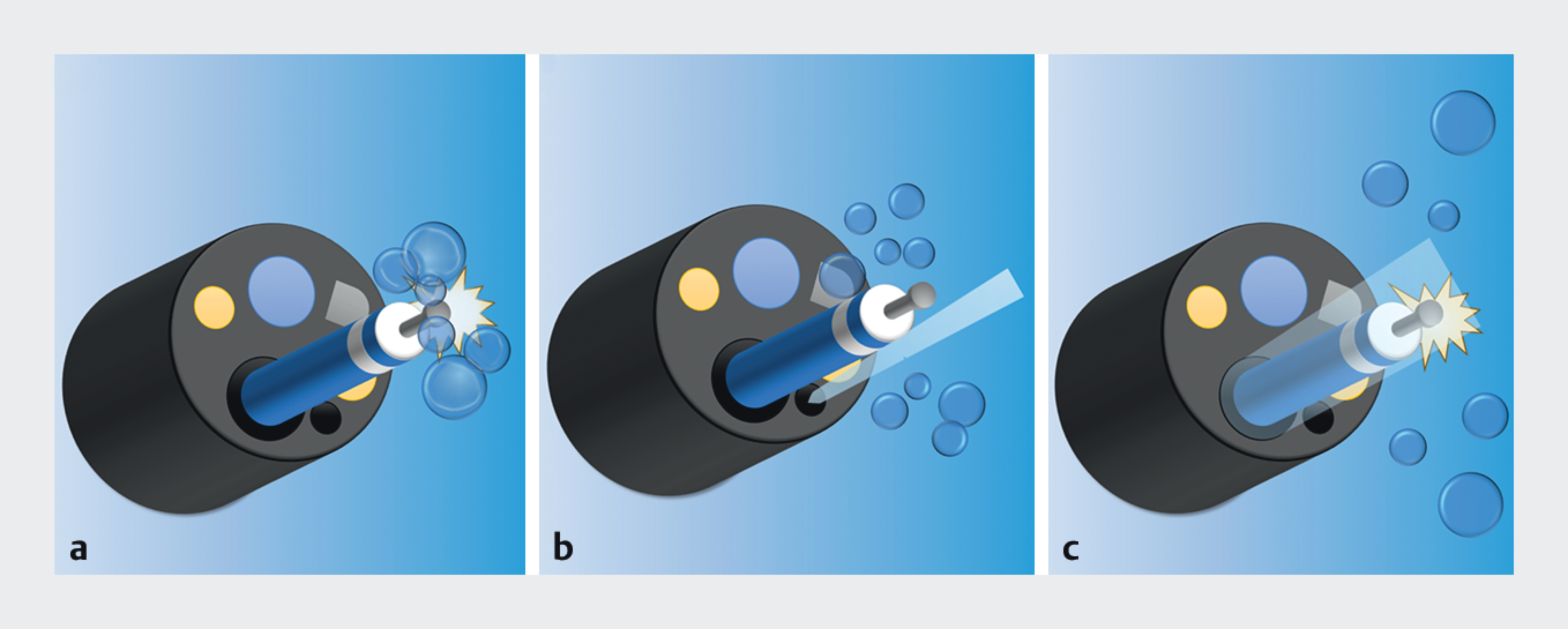
In the paired pump immersion system, the flushing pump employed by the operator connects to the endoscope’s water supply channel, whereas the flushing pump employed by the assistant connects to a forceps plug with an irrigator, allowing water delivery through the working channel ([Fig. 1], [Fig. 2]). During U-ESD, the assistant can synchronize water delivery through the working channel with the operator’s activation of the electrosurgical device to efficiently remove any bubbles ([Fig. 3]) and maintain a clear field of view ([Fig. 4], [Video 1]). Water is delivered through the working channel, which is aligned with the tip of the high-frequency device, making bubble removal more efficient than through the water supply channel alone. The assistant’s flushing pump is set to the minimum output, as excessive pressure may destabilize the field and interfere with the operator’s procedures. Therefore, the operator can focus on performing ESD without concerns regarding bubbles.


The paired pump immersion system ensures a clear field of view and improves the efficiency of U-ESD without using specialized equipment or complex setups.
Endoscopy_UCTN_Code_TTT_1AU_2AF
E-Videos is an open access online section of the journal Endoscopy, reporting on interesting cases and new techniques in gastroenterological endoscopy.
All papers include a high-quality video and are published with a Creative Commons
CC-BY license. Endoscopy E-Videos qualify for HINARI discounts and waivers and eligibility is automatically checked during the submission
process. We grant 100% waivers to articles whose corresponding authors are based in
Group A countries and 50% waivers to those who are based in Group B countries as classified
by Research4Life (see: https://www.research4life.org/access/eligibility/).
This section has its own submission website at https://mc.manuscriptcentral.com/e-videos.
Conflict of Interest
The authors declare that they have no conflict of interest.
-
References
- 1 Yahagi N, Nishizawa T, Sasaki M. et al. Water pressure method for duodenal endoscopic submucosal dissection. Endoscopy 2017; 49: E227-E228
- 2 Nagata M. Usefulness of underwater endoscopic submucosal dissection in saline solution with a monopolar knife for colorectal tumors (with videos). Gastrointest Endosc 2018; 87: 1345-1353
- 3 Abe S. Underwater colorectal endoscopic submucosal dissection: Should we be submarine voyagers?. Gastrointest Endosc 2024; 100: 1088-1089
- 4 Nomura T, Sugimoto S, Hayashi Y. et al. Colorectal endoscopic submucosal dissection using a gas-free saline-immersion dissection technique. Endoscopy 2023; 55: E1039-E1040
- 5 Sasaki M, Masunaga T, Miyazaki K. et al. Automatic water irrigation synchronized with the electrosurgical unit: Bubble-free underwater endoscopic submucosal dissection. Endoscopy 2024; 56: E468-E469
Correspondence
Publication History
Article published online:
18 July 2025
© 2025. The Author(s). This is an open access article published by Thieme under the terms of the Creative Commons Attribution License, permitting unrestricted use, distribution, and reproduction so long as the original work is properly cited. (https://creativecommons.org/licenses/by/4.0/).
Georg Thieme Verlag KG
Oswald-Hesse-Straße 50, 70469 Stuttgart, Germany
-
References
- 1 Yahagi N, Nishizawa T, Sasaki M. et al. Water pressure method for duodenal endoscopic submucosal dissection. Endoscopy 2017; 49: E227-E228
- 2 Nagata M. Usefulness of underwater endoscopic submucosal dissection in saline solution with a monopolar knife for colorectal tumors (with videos). Gastrointest Endosc 2018; 87: 1345-1353
- 3 Abe S. Underwater colorectal endoscopic submucosal dissection: Should we be submarine voyagers?. Gastrointest Endosc 2024; 100: 1088-1089
- 4 Nomura T, Sugimoto S, Hayashi Y. et al. Colorectal endoscopic submucosal dissection using a gas-free saline-immersion dissection technique. Endoscopy 2023; 55: E1039-E1040
- 5 Sasaki M, Masunaga T, Miyazaki K. et al. Automatic water irrigation synchronized with the electrosurgical unit: Bubble-free underwater endoscopic submucosal dissection. Endoscopy 2024; 56: E468-E469