

Subscribe to RSS
DOI: 10.1055/s-0045-1813705
Abducens Nerve Schwannoma: A Case Report on Extremely Rare Cranial Nerve Tumor
Authors

Abstract
Abducens nerve (CN VI) schwannomas are exceedingly rare intracranial tumors, constituting less than 10% of all primary cranial nerve schwannomas. This report details the case of a 30-year-old woman presenting with diplopia and progressive visual deterioration due to an abducens nerve schwannoma. Imaging revealed an extra-axial lesion involving the right cavernous sinus and encasing the right internal carotid artery. Surgical resection via frontotemporal craniotomy achieved near-total tumor removal, with histopathological analysis confirming a benign schwannoma. Postoperatively, the patient exhibited persistent right lateral rectus palsy as preoperative status but improvement in diplopia. This case underscores the rarity of abducens nerve schwannomas, the challenges in achieving complete resection due to neurovascular adherence, and the importance of careful surgical planning for optimal patient outcomes.
Introduction
Schwannomas, historically referred to as neurinomas and neurilemomas in the literature, account for less than 10% of all primary intracranial tumors.[1] Sensory nerves are predominantly affected; however, the optic and olfactory nerves lack a Schwann cell layer, rendering them incapable of developing schwannomas. Among the cranial nerves, the acoustic and trigeminal nerves are the most frequently involved. In contrast, schwannomas affecting other cranial nerves are rare, with those arising from purely motor nerves being exceptionally uncommon. Since the first documented case of an abducens nerve (CN VI) schwannoma reported by Chen in 1981,[2] only 31 additional cases had been reported worldwide by 2017.[3] This report presents a case of a patient experiencing diplopia followed by progressive visual deterioration in the right eye over 6 months. Neurological examination revealed right lateral rectus muscle palsy. Imaging studies identified an extra-axial lesion adjacent to the right cavernous sinus, with involvement of the cavernous sinus and encasement of the right cavernous internal carotid artery (ICA). During surgical intervention, the tumor was observed to originate from the abducens nerve, and histopathological analysis confirmed the diagnosis of schwannoma.
Case Presentation
A 30-year-old woman with no known comorbidities presented to the neurosurgery department with complaints of diplopia, followed by progressive deterioration of vision in the right eye over a duration of 6 months. On presentation, the patient was hemodynamically stable, with a pulse rate of 80 beats per minute, blood pressure of 130/70 mm Hg, and an oxygen saturation of 98% on room air. Neurological examination revealed that the patient was conscious, oriented, and able to follow verbal commands. No limb paresis was observed. Bilateral pupils measured 3 mm and were reactive to light, with a distant visual acuity of 6/6. Examination of extraocular muscle movements demonstrated right lateral rectus muscle palsy, while other ocular movements were preserved. Corneal reflex and facial sensations remained intact.
Magnetic resonance imaging (MRI) brain plain + contrast revealed an ill-defined extra-axial lesion centered around the right sphenoid wing, clinoid process, superior part of the clivus, and right cavernous sinus. The lesion measured 50 × 63 × 43 mm (anteroposterior × transverse × superoinferior) and was associated with moderate to severe surrounding edema in the right parietotemporal lobe and ganglio-capsular region, resulting in a 7-mm midline shift toward the left. The lesion involved the right cavernous sinus, right foramen ovale, and right foramen lacerum, with encasement of the right cavernous ICA (∼180 degrees) without loss of flow void. Laterally, the lesion invaded the cortex of the medial temporal lobe, while medially, it compressed the pituitary gland. MRI characteristics included T1 hypointensity, T2 fluid-attenuated inversion recovery heterogeneous hyperintensity, and heterogeneous postcontrast enhancement with nonenhancing necrotic areas. Additionally, a few areas demonstrated gradient echo blooming, with no evidence of diffusion restriction ([Fig. 1]).

The patient underwent a frontotemporal craniotomy for tumor resection. Intraoperatively, the tumor was found to originate from the abducens nerve. It was found to be adhered to the trigeminal nerve. A near-total resection was achieved, with residual tumor left behind due to adherence to critical vascular and neuronal structures over the lateral wall of the cavernous sinus. Specimen was sent for histopathological examination. The postoperative course was uneventful. The patient was extubated successfully and initially managed in the neuro intensive care unit before being transferred to the general ward. A postoperative computed tomography scan of the brain was performed ([Fig. 2]).

Histopathological examination of the excised specimen, using hematoxylin and eosin staining, demonstrated a tumor pattern characteristic of Antoni A and Antoni B regions. The Antoni A regions exhibited densely packed spindle cells with nuclear palisading and Verocay body formation, whereas the Antoni B regions displayed a hypocellular edematous stroma ([Fig. 3]). Immunohistochemical analysis revealed diffuse strong positivity for S-100 and SOX-10, with negative staining for the progesterone receptor and epithelial membrane antigen (EMA). The Ki-67 proliferative index was less than 1%, supporting the diagnosis of a benign nerve sheath tumor—schwannoma.

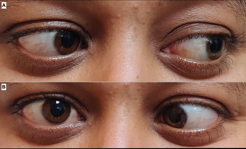
The patient's postoperative course was satisfactory, she was discharged on the fifth postoperative day with no new neurological deficits apart from the persistence of right lateral rectus palsy same as preoperative status ([Fig. 4]). She was advised to follow-up regularly for clinical and radiological evaluation. On 1-month follow-up, patient was symptomatically better in terms of resolution of diplopia. Postoperative MRI brain plain + contrast done were suggestive of very less residual lesion with significant resolution of perilesional edema ([Fig. 5]).

Discussion
Cranial nerve schwannomas originate in transitional segments where Schwann cells replace oligodendroglia to myelinate cranial nerves. The anatomical location of this transition varies among different cranial nerves.[4] In contrast to vestibular schwannomas, abducens nerve schwannomas arise at a greater distance from the glial-Schwann sheath junction, which is approximately 1 mm from the neuraxis for the abducens cranial nerve.[5]
Based on tumor location, abducens schwannomas can be classified into three types: cavernous (type 1), cisternal (type 2), and cisterno-cavernous (type 3).[6] The clinical presentation varies according to tumor location. Type 1 tumors primarily manifest as diplopia due to isolated abducens nerve palsy.[3] In contrast, cisternal tumors may present with obstructive hydrocephalus and increased intracranial pressure, in addition to abducens nerve palsy.[7] Notably, type 2 schwannomas often do not cause abducens nerve palsy, which has been attributed to the long cavernous segment of the trochlear nerve, similar to large trochlear schwannomas that rarely present with trochlear paresis.[6] The most common presentations of patients with abducens nerve schwannoma are abducens nerve palsy, followed by headache and exophthalmos.[7] Depending on tumor size, other cranial nerves, such as the trigeminal, vestibulocochlear, and facial nerves, may also be involved, leading to symptoms such as facial pain, numbness, hearing disturbances, or oculomotor palsy.[3]
The abducens nucleus is located in the pons, ventral to the floor of the fourth ventricle, slightly lateral to the midline. The paramedian pontine reticular formation is positioned medially, while the medial longitudinal fasciculus lies superior and medial to the nucleus. The fascicular portion of the abducens nerve exits ventrally, passing superior to the facial nucleus before exiting the brainstem between the pons and medulla. It then ascends along the base of the pons adjacent to the basilar artery, traverses Dorello's canal, and enters the cavernous sinus, where it is the most medial cranial nerve within the sinus. Finally, it enters the orbit through the superior orbital fissure to innervate the lateral rectus muscle.[5]
A distinct radiological feature of dumbbell-shaped schwannomas has been described, particularly in trigeminal nerve schwannomas. However, abducens nerve schwannomas exhibit a different pattern. While the neck constriction of dumbbell-shaped trigeminal schwannomas forms an obtuse angle, in abducens schwannomas, it forms an acute angle.[8]
The primary treatment for these lesions is surgical resection, with the approach depending on tumor location along the nerve tract. Cisternal schwannomas can be accessed via subtemporal, transtentorial, frontotemporal, or suboccipital approaches, allowing for gross total resection in most cases. Cavernous schwannomas are typically accessed via a frontotemporal approach, while cisterno-cavernous tumors may require either a frontotemporal or subtemporal approach. However, tumor involvement in the cavernous sinus complicates complete resection due to adherence to the ICA, often necessitating subtotal resection.[5] Maximizing tumor removal while preserving nerve function remains a key surgical objective. However, complete recovery of abducens nerve function following schwannoma resection is uncommon, as postoperative transient or permanent abducens nerve palsy frequently occurs due to nerve disturbance or sacrifice. Recovery is more likely in cases where the tumor is confined to the cavernous sinus, as the nerve within Dorello's canal is often spared.[9] Additionally, anatomical variations, such as the presence of dual nerve trunks in the cavernous segment of the abducens nerve, may contribute to the preservation of function following surgical intervention.[10] In cases where the nerve is completely transected, nerve reconstruction is recommended, with acceptable functional outcomes.[11] Among all the pathologically confirmed cases of intracranial abducens nerve schwannoma reported to date, complete postoperative nerve function recovery was achieved in only seven cases, while partial recovery was observed in three cases.[12] [13]
Imran Zaidi et al[14] reported the first case of an abducens nerve schwannoma treated via an expanded endonasal approach. This minimally invasive technique provides direct access to critical skull base structures, potentially reducing morbidity, and may be considered in patients whose tumors extend into the clivus or sphenoid sinus.
Stereotactic radiosurgery has emerged as an effective adjunct for small or residual abducens nerve schwannomas, providing durable tumor control and improvement in diplopia.[15] Apart from surgery, radiation therapy has been proposed as an adjunctive treatment for nonacoustic schwannomas, particularly in cases of subtotal resection. The rationale for its use is supported by reports demonstrating favorable outcomes in subtotally resected acoustic schwannomas treated with radiation therapy.[16]
Conclusion
Abducens nerve schwannomas are rare tumors that can present with cranial nerve deficits, most commonly diplopia due to lateral rectus muscle palsy. Accurate diagnosis requires a combination of clinical evaluation and advanced neuroimaging techniques. Surgical resection remains the primary treatment modality, with the extent of resection often limited by adherence to critical neurovascular structures. Histopathological and immunohistochemical analyses confirm the diagnosis and guide postoperative management. Despite surgical intervention, postoperative nerve dysfunction may persist, emphasizing the need for careful surgical planning and long-term follow-up. This case highlights the importance of early detection and individualized treatment strategies to optimize patient outcomes.
Conflict of Interest
None declared.
-
References
- 1 Zülch KJ. Brain Tumors: Their Biology and Pathology. 3rd ed. Berlin: Springer-Verlag; 1986
- 2 Chen BH. Neurinoma of the abducens nerve. Neurosurgery 1981; 9 (01) 64-66
- 3 Sun H, Sharma K, Kalakoti P. et al. Factors associated with abducens nerve recovery in patients undergoing surgical resection of sixth nerve schwannoma: a systematic review and case illustration. World Neurosurg 2017; 104: 883-899
- 4 Lo PA, Harper CG, Besser M. Intracavernous schwannoma of the abducens nerve: a review of the clinical features, radiology and pathology of an unusual case. J Clin Neurosci 2001; 8 (04) 357-360
- 5 Wilkins RH, Rengachary SS. Neurosurgery. 2nd ed. Vol. 2. New York: McGraw-Hill, Health Professions Division; 1996: 1553-1557
- 6 Celli P, Ferrante L, Acqui M, Mastronardi L, Fortuna A, Palma L. Neurinoma of the third, fourth, and sixth cranial nerves: a survey and report of a new fourth nerve case. Surg Neurol 1992; 38 (03) 216-224
- 7 Halalmeh DR, Asad MY, Atallah OA. et al. Predictors of surgical outcomes in patients with abducens nerve schwannoma: a comprehensive systematic review and a case report. World Neurosurg 2023; 179: 204-215.e4
- 8 Shibao S, Hayashi S, Yoshida K. Dumbbell-shaped abducens schwannoma: case report. Neurol Med Chir (Tokyo) 2014; 54 (04) 331-336
- 9 Nakagawa T, Uchida K, Ozveren MF, Kawase T. Abducens schwannoma inside the cavernous sinus proper: case report. Surg Neurol 2004; 61 (06) 559-563 , discussion 563
- 10 Haładaj R, Skrzat J. Bilateral duplication of the abducens nerve - case study. Folia Med Cracov 2019; 59 (04) 13-20
- 11 Sekhar LN, Lanzino G, Sen CN, Pomonis S. Reconstruction of the third through sixth cranial nerves during cavernous sinus surgery. J Neurosurg 1992; 76 (06) 935-943
- 12 Vachata P, Sameš M. Abducens nerve schwannoma mimicking intrinsic brainstem tumor. Acta Neurochir (Wien) 2009; 151 (10) 1281-1287
- 13 Nakamura M, Carvalho GA, Samii M. Abducens nerve schwannoma: a case report and review of the literature. Surg Neurol 2002; 57 (03) 183-188 discussion 188–189
- 14 Imran Zaidi SM, Naik P, Ahmed SK. Abducens nerve schwannoma: first case to be treated with an expanded endonasal approach. Ear Nose Throat J 2022; 101 (05) NP190-NP192
- 15 Langlois AM, Iorio-Morin C, Faramand A. et al. Outcomes after stereotactic radiosurgery for schwannomas of the oculomotor, trochlear, and abducens nerves. J Neurosurg 2021; 135 (04) 1044-1050
- 16 Pollock BE, Kondziolka D, Flickinger JC, Maitz A, Lunsford LD. Preservation of cranial nerve function after radiosurgery for nonacoustic schwannomas. Neurosurgery 1993; 33 (04) 597-601
Address for correspondence
Publication History
Article published online:
05 January 2026
© 2026. Asian Congress of Neurological Surgeons. This is an open access article published by Thieme under the terms of the Creative Commons Attribution-NonDerivative-NonCommercial License, permitting copying and reproduction so long as the original work is given appropriate credit. Contents may not be used for commercial purposes, or adapted, remixed, transformed or built upon. (https://creativecommons.org/licenses/by-nc-nd/4.0/)
Thieme Medical and Scientific Publishers Pvt. Ltd.
A-12, 2nd Floor, Sector 2, Noida-201301 UP, India
-
References
- 1 Zülch KJ. Brain Tumors: Their Biology and Pathology. 3rd ed. Berlin: Springer-Verlag; 1986
- 2 Chen BH. Neurinoma of the abducens nerve. Neurosurgery 1981; 9 (01) 64-66
- 3 Sun H, Sharma K, Kalakoti P. et al. Factors associated with abducens nerve recovery in patients undergoing surgical resection of sixth nerve schwannoma: a systematic review and case illustration. World Neurosurg 2017; 104: 883-899
- 4 Lo PA, Harper CG, Besser M. Intracavernous schwannoma of the abducens nerve: a review of the clinical features, radiology and pathology of an unusual case. J Clin Neurosci 2001; 8 (04) 357-360
- 5 Wilkins RH, Rengachary SS. Neurosurgery. 2nd ed. Vol. 2. New York: McGraw-Hill, Health Professions Division; 1996: 1553-1557
- 6 Celli P, Ferrante L, Acqui M, Mastronardi L, Fortuna A, Palma L. Neurinoma of the third, fourth, and sixth cranial nerves: a survey and report of a new fourth nerve case. Surg Neurol 1992; 38 (03) 216-224
- 7 Halalmeh DR, Asad MY, Atallah OA. et al. Predictors of surgical outcomes in patients with abducens nerve schwannoma: a comprehensive systematic review and a case report. World Neurosurg 2023; 179: 204-215.e4
- 8 Shibao S, Hayashi S, Yoshida K. Dumbbell-shaped abducens schwannoma: case report. Neurol Med Chir (Tokyo) 2014; 54 (04) 331-336
- 9 Nakagawa T, Uchida K, Ozveren MF, Kawase T. Abducens schwannoma inside the cavernous sinus proper: case report. Surg Neurol 2004; 61 (06) 559-563 , discussion 563
- 10 Haładaj R, Skrzat J. Bilateral duplication of the abducens nerve - case study. Folia Med Cracov 2019; 59 (04) 13-20
- 11 Sekhar LN, Lanzino G, Sen CN, Pomonis S. Reconstruction of the third through sixth cranial nerves during cavernous sinus surgery. J Neurosurg 1992; 76 (06) 935-943
- 12 Vachata P, Sameš M. Abducens nerve schwannoma mimicking intrinsic brainstem tumor. Acta Neurochir (Wien) 2009; 151 (10) 1281-1287
- 13 Nakamura M, Carvalho GA, Samii M. Abducens nerve schwannoma: a case report and review of the literature. Surg Neurol 2002; 57 (03) 183-188 discussion 188–189
- 14 Imran Zaidi SM, Naik P, Ahmed SK. Abducens nerve schwannoma: first case to be treated with an expanded endonasal approach. Ear Nose Throat J 2022; 101 (05) NP190-NP192
- 15 Langlois AM, Iorio-Morin C, Faramand A. et al. Outcomes after stereotactic radiosurgery for schwannomas of the oculomotor, trochlear, and abducens nerves. J Neurosurg 2021; 135 (04) 1044-1050
- 16 Pollock BE, Kondziolka D, Flickinger JC, Maitz A, Lunsford LD. Preservation of cranial nerve function after radiosurgery for nonacoustic schwannomas. Neurosurgery 1993; 33 (04) 597-601