

Subscribe to RSS
DOI: 10.1055/s-0044-1788895
Multiple Free Flaps and Second Toe Transfer to Salvage Grasp Function in Bilateral Complete Degloved Hands
Authors
Funding None.


Abstract
A 21-year-old male laborer sustained bilateral degloving injury of the hands with multiple digital amputations and devascularized digits. After X-rays, preliminary debridement was done, when digital amputations were completed, including index ray amputation on both sides. The next day, two anterolateral thigh (ALT) flaps and one second toe transfer were done to restore coverage in the palm and the web and reconstruct the lost thumb. This ensured both coverage and thumb length on the right side, but on the left side the procedure was terminated with flap transfer only. After 3 months, the other second toe was harvested with a dorsalis pedis flap for reconstruction of the left thumb, and a free gracilis flap was done for optimal donor site coverage in the donor left foot. Evaluation after 2 years showed functional hands with reasonable power grasp, pinch grip, and dexterity to manipulate small and large objects that permitted an independent living.
Introduction
Degloving injuries of the whole hand with amputation of one or more digits are problems that need good planning to not only provide good skin coverage but also restore mobile digits to enable prehension. The condition is even more critical when both hands are injured.
Only flap covers impel a second surgical procedure to make the phalangeal or metacarpal hand functional. Traditional methods like first web deepening and distraction lengthening of short digital stumps can improve hand function, but a toe transfer is an elegant method to achieve the same objective.
Li et al[1] report combined deep inferior epigastric perforator (DIEP) and second toe transfer in five cases, done either electively or as a delayed emergency; the DIEP flap was joined to the radial vessels end to side proximal to the site of anastomosis of the transferred second toe.
This is a case report of a bilateral circumferential degloving injury of the hands wherein simultaneous free flap cover and second toe transfer were planned for both sides. One of the toe transfers had to be scheduled for second-stage surgery in the interest of patient safety.
Surgical Technique
Stage 1
Debridement of both hands was done within 6 hours after the trauma. The degloved skin was distally based at the proximal phalanx level with amputation of the middle and distal phalanges for the fingers. Since the skin was devascularized and crushed, it was removed. The patient underwent transphalangeal amputation of fourth and fifth digits. The middle finger was amputated proximal to the metacarpo phalangeal joint (MPJ) on both sides. The thumb amputations left a stump at the base of the proximal phalanx. A ray amputation of both index fingers was done to widen the first webspace. Loss of skin extended up to the wrist crease.
The next day (12 hours after the debridement), a semi-elective reconstruction was planned to transfer two anterolateral thigh (ALT) flaps and two second toe transfers to cover the degloved hand and to lengthen the amputated thumbs on both sides.
Attention was first directed to reconstructing the right hand. The ALT flap was harvested from the left thigh and simultaneously the second toe from the left foot. Revascularization of the ALT was completed first before the second toe was detached. Anastomosis details are presented in [Table 1].
|
Right hand |
Left hand |
Anastomosis artery |
Anastomosis vein |
|||
|---|---|---|---|---|---|---|
|
Flap |
Toe |
Flap |
||||
|
Stage 1 |
Left ALT |
Left second toe |
Right ALT flap[a] |
Flap artery: radial artery end to end Toe artery: descending branch of the ALT flap end to end |
Flap vein: tributary of the cephalic vein Toe vein: deep vein to the radial venae comitantes Saphenous vein to the cephalic vein |
|
|
Flap |
Toe |
|||||
|
Stage 2 |
Gracilis muscle |
Right second toe + dorsalis pedis flap |
Toe + dorsalis pedis: radial artery end to side proximal to a previous ALT flap anastomosis Gracilis: anterior tibial artery proximal to the site of detachment of the donor toe dorsalis pedis flap |
Toe + flap vein: deep vein to the radial venae comitantes Saphenous vein to the rerouted basilic vein Gracilis: 2 venae comitantes of the anterior tibial artery |
||
Abbreviation: ALT, anterolateral thigh.
a The second toe transfer was deferred for the left hand on account of increasing operative duration.
Once the left second toe donor site was closed primarily, the second ALT flap was harvested from the right thigh. Since the operative duration now had crossed 6 hours, the harvest of the right second toe for the left hand was deferred and the procedure ended with transfer of the ALT flap only to the left hand (∼9 hours of operative time; [Figs. 1] and [2]).


All the microvascular anastomoses were successful in the immediate post-op period. Both the ALT flap donor sites needed split skin grafts.
Stage 2
Eight weeks later, a second toe transfer from the right foot was done for the thumb reconstruction in the left hand. Since a difficulty was anticipated in the availability of existing ALT flap at the recipient site, the toe was harvested in continuity with a dorsalis pedis flap. Anastomosis was done by harvesting the pedicle of the toe up to the anterior tibial vessels just above the ankle to anastomose to the same radial artery and accompanying veins proximal to the previous anastomotic site in the upper limb ([Figs. 3] and [4]).


A gracilis flap was harvested from the left thigh and anastomosed to the stump of the anterior tibial vessels above the left ankle to ensure good wound healing in the foot. Both the transferred flaps survived uneventfully.
K-wires used to fix the second toes that were removed between the fourth and sixth weeks postoperatively and therapy was instituted. One sitting of flap thinning was done on the right side.
Results
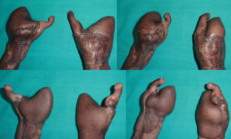
The aim was to provide a mobile thumb of reasonable length to oppose the phalangeal ulnar aspect of either hand. The patient was able to grasp using both hands simultaneously. He could use the tip of the reconstructed thumb for fine activities and had a strong adduction of the thumb against the third metacarpal. The available active range of motion (ROM) of the reconstructed thumb was on average 30 degrees at the basal joint, 20 degrees at the MPJ and 20 degrees at the interphalangeal joint (IPJ). He had a moving 2-point discrimination (2PD) of between 7 and 9 mm. There were no trophic ulcerations in spite of activities of daily living. Using an activity board to document the possible activities of daily living emphasized the functionality of the reconstruction.
In spite of the flap being insensate, there was no evidence of any trophic ulcers in the hand. A thin flap was felt to be more appropriate to give a better appearance ([Figs. 5] and [6]).

Discussion
Zelken et al[2] described the successful use of combined ALT and groin pedicle flaps for the total degloved hand. An average of one to four (mean: 1.8) revision procedures were needed to improve appearance and function and only one case needed a great toe wraparound subsequently to lengthen the thumb.
Lin[3] further analyzed 15 cases of degloved hands, of which 4 were total degloving covered with pedicle or free groin or ALT flap. Timely division of local flap and syndactyly separation with simultaneous thinning permitted earlier rehabilitation. The amputations were not significant to warrant reconstruction.
Kim et al[4] report a single DIEP flap used for total degloving hand with thumb amputation at the MPJ. Images show all digits with stumps at the level of the proximal phalanx, but no reconstruction was offered.
In the present study, the bilateral nature of the injury and amputations that compromised the number of digits and/or the length made it more imperative to offer primary reconstruction.
Thomas and Tsai[5] describe double toe transfers for reconstructing the middle and ring fingers in combination with a distally based radial forearm flap, using the stump of the radial artery for revascularization of the transferred toes. The thumb was uninjured.
Primary toe transfer makes optimal use of recipient pedicles, enabling earlier rehabilitation to permit good, mobile digits.
Fan et al[6] in 2006 reported the largest series of 11 cases of pure thumb loss with a soft-tissue defect, wherein the scapular or the ALT flap provided coverage and the second or first toe wraparound. The anastomoses used either the radial artery for the toe and the deep plantar branch or the plantar arch for flap perfusion.
We used the descending branch of the ALT flap for toe perfusion in one hand. Since the other toe transfer was done in a second stage, it was done end to side to the radial artery at a higher level.
We planned to provide a mobile radial column (the thumb) of adequate length so that it could oppose the ulnar post consisting of the fourth and fifth digits. These had been amputated distal to the MPJ. Hence, the flexion afforded by the MPJ and the natural mobility of the fourth and fifth carpometacarpal joint was considered enough to provide grasp. The thumb could also be positioned to permit finer activities like manipulating tools including the use of a cellphone.
One problem that we faced with the reconstruction on the left hand where a simultaneous toe transfer was not done was a relative lack of normal ALT flap skin (on account of contraction) that forced a modification, leading to the elevation of the dorsalis pedis flap in continuity.
To avoid inadequate split skin graft on the foot donor site following flap elevation, a free gracilis flap was transferred simultaneously as a simple option.
In Shen et al,[7] in 3 of 31 cases with degloving and multiple digital losses, they used either two great toes or one great toe and one double toe transfer to the same hand for finger losses, and stressed on the importance of radial digital length for good function.
Combined toe transfers or great toe transfers result in substantial donor defects, which may not be acceptable for Indian patients who rely on open-toed footwear. To minimize donor morbidity, the described method was adopted, and the patient can use Indian-type footwears with ease.
Our intention was to achieve a reasonably functional hand by using a procedure with minimum donor site sequelae. At the end of follow-up, the patient's Michigan Hand Outcomes Questionnaires (MHQ) score was 63.97 for the right hand, 60.64 for the left hand, and 62.30 for both hands together ([Videos 1] and [2]).
Video 1 This video shows reasonable achievement of normal daily activities and bimanual activities at the 2-year follow-up.
Video 2 This video shows reasonable achievement of normal daily activities and bimanual activities at the 2-year follow-up.
Conclusion
Simultaneous toe transfer and free flap coverage optimize digital mobility and wound closure in degloving hand injuries with digital amputations. When the logistics of operative time do not permit transfer of tissues in a single stage in a bilateral injury, early planned second-stage reconstruction is crucial to restore activities of daily living.
Conflict of Interest
None declared.
-
References
- 1 Li XJ, Tong J, Wang Y. Combined free toe and free deep inferior epigastric perforator flap for reconstruction of the thumb and thumb web space. J Reconstr Microsurg 2000; 16 (06) 427-436
- 2 Zelken JA, Chang NJ, Wei FC, Lin CH. The combined ALT-groin flap for the mutilated and degloved hand. Injury 2015; 46 (08) 1591-1596
- 3 Lin TS. One-stage debulking procedure after flap reconstruction for degloving injury of the hand. J Plast Reconstr Aesthet Surg 2016; 69 (05) 646-651
- 4 Kim MJ, Park BY. From mitten hand to five individual fingers: one-stage coverage using a deep inferior epigastric perforator free flap for an extensive degloving hand injury. Journal of Reconstructive Microsurgery Open 2018; 03: e28-e31
- 5 Thomas BP, Tsai TM. Primary reconstruction of a degloved hand using multiple toe transfers on a single pedicle and a reversed radial artery flap. J Reconstr Microsurg 2004; 20 (01) 3-6
- 6 Fan C-Y, Jiang J, Zeng B-F, Jiang P-Z, Cai P-H, Chung KC. Reconstruction of thumb loss complicated by skin defects in the thumb-index web space by combined transplantation of free tissues. J Hand Surg Am 2006; 31 (02) 236-241
- 7 Shen XF, Mi JY, Xue MY. et al. Modified great toe wraparound flap with preservation of plantar triangular flap for reconstruction of degloving injuries of the thumb and fingers: long-term follow-up. Plast Reconstr Surg 2016; 138 (01) 155-163
Address for correspondence
Publication History
Article published online:
05 August 2024
© 2024. Association of Plastic Surgeons of India. This is an open access article published by Thieme under the terms of the Creative Commons Attribution-NonDerivative-NonCommercial License, permitting copying and reproduction so long as the original work is given appropriate credit. Contents may not be used for commercial purposes, or adapted, remixed, transformed or built upon. (https://creativecommons.org/licenses/by-nc-nd/4.0/)
Thieme Medical and Scientific Publishers Pvt. Ltd.
A-12, 2nd Floor, Sector 2, Noida-201301 UP, India
-
References
- 1 Li XJ, Tong J, Wang Y. Combined free toe and free deep inferior epigastric perforator flap for reconstruction of the thumb and thumb web space. J Reconstr Microsurg 2000; 16 (06) 427-436
- 2 Zelken JA, Chang NJ, Wei FC, Lin CH. The combined ALT-groin flap for the mutilated and degloved hand. Injury 2015; 46 (08) 1591-1596
- 3 Lin TS. One-stage debulking procedure after flap reconstruction for degloving injury of the hand. J Plast Reconstr Aesthet Surg 2016; 69 (05) 646-651
- 4 Kim MJ, Park BY. From mitten hand to five individual fingers: one-stage coverage using a deep inferior epigastric perforator free flap for an extensive degloving hand injury. Journal of Reconstructive Microsurgery Open 2018; 03: e28-e31
- 5 Thomas BP, Tsai TM. Primary reconstruction of a degloved hand using multiple toe transfers on a single pedicle and a reversed radial artery flap. J Reconstr Microsurg 2004; 20 (01) 3-6
- 6 Fan C-Y, Jiang J, Zeng B-F, Jiang P-Z, Cai P-H, Chung KC. Reconstruction of thumb loss complicated by skin defects in the thumb-index web space by combined transplantation of free tissues. J Hand Surg Am 2006; 31 (02) 236-241
- 7 Shen XF, Mi JY, Xue MY. et al. Modified great toe wraparound flap with preservation of plantar triangular flap for reconstruction of degloving injuries of the thumb and fingers: long-term follow-up. Plast Reconstr Surg 2016; 138 (01) 155-163