

Subscribe to RSS
DOI: 10.1055/s-0043-1771406
Magnetic Resonance Imaging Spectrum of Cauda Equina and Conus Lymphoma: Keys to Unravel the Differential Diagnosis with a Review of the Literature
Authors

Abstract
Central nervous system lymphoma is not an uncommon condition, but spinal lymphoma with isolated involvement of the conus medullaris and cauda equina is a rare entity. Our study aims to evaluate the various magnetic resonance imaging (MRI) features of cauda equina and conus lymphoma. This retrospective study was carried out on nine patients with histopathologically proven cauda equina and conus lymphoma, who were presented at a tertiary care hospital between January 2018 and June 2020. All patients underwent lumbar spine MRI scans using a 1.5 Tesla MR scanner. The clinical data and different MRI findings were analyzed with an independent sample t-test and paired-samples t-test. Among the nine patients with cauda equina and conus lymphoma, three had primary lymphoma and six had secondary lymphoma. Six patients (66.7%) showed a diffuse pattern of involvement of cauda equina and conus medullaris, while three patients (33.3%) showed a focal pattern. T2-weighted imaging (T2WI) hypo to isointense signal intensity lesions were observed in six patients (66.7%) and T2WI iso to slight hyperintensities in three patients (33.3%). Diffuse sheet-like thickening and postcontrast enhancement of the thickened cauda equina nerve roots were observed in two patients of primary and one patient with secondary lymphoma. The diagnosis of cauda equina and conus lymphoma especially primary lymphoma is challenging and requires a high index of clinical suspicion as distinguishing this entity from similar conditions is difficult solely on MRI. Early diagnosis of this entity is important for early institution of treatment for increasing the chances of survival and improvement of symptoms.
Keywords
non-Hodgkin lymphoma - magnetic resonance imaging (MRI) - cauda equina - diffusion-weighted imaging - chemoradiotherapyIntroduction
The intradural and intramedullary spinal lymphomas, whether primary or secondary, have been an uncommon entity. Central nervous system (CNS) lymphoma is rather common but spinal lymphoma is a rare entity.[1] Primary CNS lymphoma accounts for 1 to 2% of all non-Hodgkin lymphoma.[2] Of the CNS lymphoma, spinal cord lymphoma occurs in less than 1%.[3] Among the spinal cord lymphoma, cauda equina lymphoma is the rarest one.[3]
The primary cauda equina and conus lymphomas are uncommon extranodal non-Hodgkin lymphoma. Secondary cauda equina and conus lymphomas occur due to secondary involvement of systemic lymphoma, either from hematogenous, direct invasion, venous spread through Batson's plexus, or drop metastasis from brain involvement.[4]
B cell type of lymphoma dominantly affects the cauda equina in both primary and secondary forms.[2] The aggressive form of B cell lymphoma secondarily affects the CNS in up to 25% of patients. Human immunodeficiency virus (HIV)-infected, organ-transplanted, and congenital immunodeficiency patients are more prone to the development of CNS lymphoma.[5] There has been an increasing incidence of primary CNS lymphoma in immune-competent individuals also. The frequency of spinal lymphomas in decreasing order are intraosseous, epidural, intradural extramedullary, and intramedullary depending on their location.[6]
Leptomeningeal seeding of lymphoma manifests as abnormal shaggy to nodular enhancement over the spinal cord surface and nerve roots.[4] The intravascular form of B cell lymphoma presents with progressive cauda equina syndrome, skin lesions, or other systemic features[7] where nasal mucosal, skin, or bone marrow biopsy helps in its diagnosis.
Intraosseous and epidural lymphomas have a better prognosis than intradural or intramedullary lymphoma,[4] so knowledge of various magnetic resonance imaging (MRI) appearances of intradural lymphoma of cauda equina and conus medullaris is of utmost importance for early diagnosis and early treatment for better patient outcomes.
This study aims to evaluate the various MRI features of cauda equina and conus lymphoma.
Materials and Methods
A retrospective study was conducted in our institute on histopathologically proven nine patients of cauda equina and conus lymphoma presented between January 2018 and June 2020. This study was approved by the institutional ethics review committee.
Inclusion Criteria
-
Only intradural extramedullary and intramedullary forms of spinal lymphoma affecting cauda equina and conus medullaris.
Exclusion Criteria
-
Isolated spinal epidural lymphoma
-
Lymphoma involving contiguous vertebra and or retroperitoneum
MRI Protocols
All patients underwent an MRI scan of the spine, using a 1.5 Tesla MR scanner, Siemens Magnatom Avanto (Siemens Medical Systems, Erlangen, Germany). The conventional MRI sequences of the spine includes sagittal T1-weighted imaging (T1WI), T2WI, short tau inversion recovery (STIR), diffusion-weighted imaging, coronal STIR, and axial T1WI, T2WI. Postgadolinium T1WI sequences were obtained in all three planes. The parameters of the various sequences used are shown in ([Table 1]).
Abbreviations: DWI, diffusion-weighted imaging; MRI, magnetic resonance imaging; STIR, short tau inversion recovery; T1WI, T1-weighted imaging; TE, time of echo; TI, inversion time; TR, repetition time.
MRI evaluation : The patients were evaluated for the involvement of conus medullaris, cauda equina nerve roots, superior or inferior extensions, and pattern of contrast enhancement.
Laboratory test and histopathological examination : All patients were evaluated with complete blood count, enzyme-linked immunosorbent assay for HIV, cerebrospinal fluid (CSF) analysis, bone marrow aspiration cytology, and posteroanterior view of chest X-rays. Confirmation of lymphoma was established by histological confirmation of postoperative resected specimen of thickened cauda equina nerve roots in three patients of primary Lymphoma, lymph nodal biopsy in five patients, and associated leg mass biopsy in one patient with secondary lymphoma.
Treatment: Four patients were treated with only multidrug chemotherapy and five patients with combined chemoradiotherapy. Clinical, hematological, or radiological follow-up was done in all patients.
Statistical analysis: All statistical analysis was performed by using Statistical Package for the Social Science (SPSS programs, version 16). The clinical data and different MRI findings were analyzed with an independent sample t-test and paired-samples t-test.
Results
Patient and clinical data: In this study sample, nine patients (n = 4 male, n = 5 female) of cauda equina and conus lymphoma were included with a male: female ratio of 1:1.25 and mean age of 48.89 ± 1.84 (standard deviation, SD) years (range: 18–80 years). The demography, clinical and MRI findings were summarized in ([Table 2]). Primary lymphoma was identified in three patients (33.3%) and secondary lymphoma in six patients (66.7%). Patients with secondary lymphoma had cervical lymphadenopathy in two patients, mediastinal lymphadenopathy in two patients, retroperitoneal lymphadenopathy in one patient, and leg mass with tibial involvement in another one patient.
Abbreviations: CSF, cerebrospinal fluid; DWI, diffusion-weighted imaging; LBA, low back ache; MRI, magnetic resonance imaging; T2WI, T2-weighted imaging; TLC, total leuckocyte count.
The most common clinical presentation was chronic paraparesis with bladder and bowel dysfunction in five patients (55.56%), chronic low backache with radiculopathy in two patients (22.2%), and chronic low backache with dissociative anesthesia in one patient (11.1%) and chronic low backache in another 1 patient (11.1%). The mean duration between neurological symptoms onset and MRI examination was 4.78 ± 2.11 (SD) months.
MRI findings: Six patients (66.7%) showed a diffuse pattern ([Figs. 1] and [2]) and three patients (33.3%) showed a focal pattern of involvement of cauda equina and conus medullaris ([Figs. 3] [4] [5]). T2WI hypo to isointense signal intensities were observed in six patients (66.7%) and T2 iso to slight hyperintensities in three patients (33.3%). Diffusion restriction with a low apparent diffusion coefficient value was observed in four patients (44.4%) with the diffuse pattern of lymphoma and two patients (22.2%) with focal lymphoma.
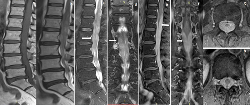


Diffuse sheet-like postcontrast enhancement of the cauda equina fibers and sheet-like thickened nerve roots was observed in two patients (22.2%) of primary ([Figs. 1] and [2]) and one patient (11.1%) with secondary lymphoma. Plaque to focal mass-like postcontrast enhancement within or around the conus medullaris and cauda equina fibers was found in four patients (44.4%) ([Figs. 3] and [5]), focal (intradural mass-like) postcontrast enhancement over conus in one patient (11.1%) ([Fig. 4]), and isolated conus surface enhancement in another one patient (11.1%).
Clumping of cauda equina nerve roots was observed in six patients (66.7%) ([Figs. 1] and [2]). Intramedullary enhancing conus lesion observed in one patient (11.1%) ([Fig. 5]), sheet-like conus surface enhancement in four patients (44.4%), and both intramedullary lesion and conus surface enhancement in four patients (44.4%) ([Fig. 3]). The salient differences between the primary and secondary lymphoma in our study sample were shown in [Table 3].
Abbreviations: DWI, diffusion-weighted imaging; MRI, magnetic resonance imaging; SD, standard deviation.
Four patients of conus and cauda equina lymphoma were treated with only multidrug chemotherapy and five patients with combined chemoradiotherapy. The mean patient survival was 6.67 ± 2.55 (SD) months after the treatment.
Discussion
Previous literature showed various case reports of lymphomatous involvement of conus and cauda equina in the localized[8] as well as a disseminated form of non-Hodgkin lymphoma. Recurrent lymphoma of spinal cord can cause cauda equina syndrome.[9] The intramedullary form of lymphoma commonly affects the thoracic cord followed by the conus medullaris and cervical cord.[10]
Primary adrenal lymphoma may be presented with cauda equina syndrome with secondary deposits in cauda equina nerve roots.[11] Patients with cauda equina lymphoma usually presented with nonspecific symptoms like low backache, radiculopathy, paraparesis, or saddle anesthesia.[12] [13]
Increased CSF protein level, hypoglycemia, or pleocytosis was observed in cauda equina lymphoma.[11] Lachance et al[14] found malignant lymphocytes in CSF in 66% of patients with cauda equina lymphoma. Accurate diagnosis of cauda equina lymphoma is usually achievable with surgical resection of the affected nerve root and confirmation on histopathological or immune histopathological examinations.[15]
Cauda equina lymphoma on MRI appears as a focal increase in the volume of cauda equina or nerve roots, sheet-like thickening, and homogenous moderate-to-intense postcontrast enhancement of the thickened nerve roots.[8] [12] In our study, sample of 66.7% of patients showed a diffuse pattern and 33.3% showed a focal pattern of involvement of cauda equina and conus medullaris. In our study sample, diffuse sheet-like postcontrast enhancement of the cauda equina fibers and sheet-like thickened nerve roots was observed in 33.3% of patients, while previous case reports by Broen et al[13] and Biasi et al[16] showed similar findings of cauda equina lymphoma. A few previous case reports by Teo et al,[3] Nakashima et al,[12] and Shin et al[17] showed focal intradural mass lesions around the conus medullaris and cauda equina but in our study sample, only 1 patient showed an intradural mass.
Focal plaque-like enhancing cauda equina mass can be encountered in cauda equina lymphoma and in such a situation tumors like schwannoma, ependymoma, neurofibroma, meningioma, dermoid, epidermoid, paraganglioma, hemangioblastoma and metastasis needed to be differentiated with their characteristics MRI features.[12] Usually, schwannoma shows T1W isointense and T2W hyperintensity with irregular postcontrast enhancement with or without cystic degeneration and commonly having neural exit foraminal extension. Meningioma showed T1W isointense and T2W iso to hypointense signal intensity with moderate to intense homogenous postcontrast enhancement and the presence of a dural tail. Ependymoma showed T1W isointense and T2W hyperintense signal intensity with intense postcontrast enhancement with or without intratumoral hemorrhage and cystic changes. Neurofibroma shows the dumbbell shape of the mass with neural exit foraminal extension.
Sheet-like cauda equina nerve root thickenings can be encountered in acute inflammatory demyelinating polyradiculoneuropathy, chronic inflammatory demyelinating polyradiculoneuropathy, Landry-Guillain-Barre syndrome, hereditary sensory-motor neuropathies (Charcot-Marie Tooth disease), sarcoidosis, arachnoiditis, tuberculous pachymeningitis, schistosomiasis,[8] [11] [18] tumor with CSF drop metastasis (glioblastoma, ependymoma, medulloblastoma, ependymoma, pineal tumor), and extracranial metastasis from breast and lung cancers.[18] [19] There is difficulty in distinguishing these conditions from cauda equina lymphoma on MRI. Even though few salient MRI findings were shown in [Table 4] for their differentiation.
|
Parameter |
Conus and cauda equina lymphoma |
Leptomeningeal metastasis |
Chronic inflammatory demyelinating polyradiculoneuropathy (CIDP) |
Neurosarcoidosis |
Inflammatory arachnoiditis |
Guillain–Barre syndrome |
|---|---|---|---|---|---|---|
|
Probable etiology |
-Most commonly non-Hodgkin lymphoma |
-Hematogenous metastasis -CSF drop metastasis from brain tumors.[23] -Extracranial metastasis (from breast and lung cancers) |
-Neurological disorder of peripheral nerve causing peripheral neuropathy.[24] -Demyelination of peripheral nerves |
-Systemic granulomatous disease[25] -Noncaseating granulomas |
-Infectious meningitis -Postoperative failed back -Postirradiation -Hemorrhage -Degenerative spondylosis |
-Acute inflammation demyelinating polyradiculoneuropathy -Postinfectious/ postvaccinal demyelination |
|
Salient MRI features |
Various MRI patterns can be seen[11] [17] [19] -Enlarged cauda equina with iso to slight hyper or hypointensities on T1WI and T2WI -Focal or diffuse homogenous enhancement over conus and cauda equina nerve roots -Diffuse sheet like postcontrast enhancement of the cauda equina nerve roots |
4 MRI pattern can be seen[26] -Solitary focal at bottom of thecal sac or along cord surface -Diffuse sheet-like coating of spinal cord/ nerve roots -Rope-like thickening of cauda equina nerves -Multifocal, discrete nodules along the spinal cord/ nerve roots |
-Enhancement of the cauda equina nerve roots -Affected nerve roots are enlarged -Most commonly enlarged in extra- foraminal segment[27] |
Smooth postcontrast enhancement over conus surface and cauda equina nerve roots[28] |
3 MRI patterns cauda nerve roots[29] - Central clumping - Peripheral clumping with empty sac sign -Central mass-like clumping with decreased thecal sac diameter |
-Ventral nerve roots dominantly affected -Normal on T1W and T2W images -Smooth enhancement over conus and cauda equina nerve roots |
Abbreviations: CSF, cerebrospinal fluid; MRI, magnetic resonance imaging; T1WI, T1-weighted imaging.
Most of patients with cauda equina lymphoma usually respond to chemotherapy, radiotherapy or combined chemoradiotherapy. In treated cauda equina lymphoma patients, the common symptoms like pain and motor weakness are usually relieved after treatment; however, bladder and bowel incontinence usually remain.[3] [20] Combined chemotherapy and radiotherapy had survival rates ranging from 16 to 44.5 months in cauda equina lymphoma patients[3] however, in our study sample, the survival period is less at 6.67 ± 2.55 (SD) months.
However, combined chemoradiotherapy is more effective than isolated radiotherapy or chemotherapy in the management of primary CNS lymphoma.[21] Isolated radiotherapy is initially effective, but its response is short-lived.[21] Aggressive surgical resection is not effective in primary CNS lymphoma.[21] But treatment protocol including initial radiotherapy or chemotherapy with or without surgical intervention is the mainstay of treatment in cauda equina lymphoma presenting with radiculopathy[22] [Table 5] shows the literature review of cauda equina and conus lymphoma in the last decade.
|
Series/year |
Number |
Age/mean age (y) |
Sex/sex ratio |
Salient MRI findings |
Form of lymphoma |
Cell type of lymphoma |
Treatment |
Outcome |
|---|---|---|---|---|---|---|---|---|
|
Iwasaki et al 2012[27] |
1 |
69 |
M |
Enhancing lesion in conus medullaris and cauda equina with old collapse of L1 vertebra |
Primary |
DLBCL |
Chemotherapy (MTX) + RT |
Initially improved but died after 18 months |
|
Biasi et al 2015[16] |
1 |
67 |
F |
Thickening of cauda equina nerve roots with intense postcontrast enhancement |
Primary |
DLBCL |
Chemotherapy |
Improved |
|
Nishida et al 2012[20] |
1 |
47 |
M |
Swelling of cauda equina with marked diffuse enhancement |
Primary |
DLBCL |
Chemotherapy +RT |
Improved |
|
Broen et al 2014[13] |
2 |
75,71 |
F:2 |
Enhancement along cauda equina fibers in first case. Thickening and enhancement of multiple lumbosacral nerves in second case |
Primary |
DLBCL |
First case- steroid second case—chemotherapy |
First case –not improved and died Second case improved |
|
Nakashima et al 2014[12] |
1 |
59 |
M |
Intradural lesion from D12 to S1 level |
Primary |
DLBCL |
RT and MTX |
Improved |
|
Teo et al 2012[3] |
1 |
58 |
M |
Minimally enhancing intradural mass from D12 to L4 level |
Primary |
DLBCL |
Chemotherapy + RT+ steroid |
Improved |
|
Shin et al 2015[17] |
1 |
79 |
F |
Segmental intradural mass from L3 to L5 level with leptomeningeal enhancement over cord and Conus. |
Primary |
DLBCL |
Chemotherapy +RT |
Improved |
|
Ogilvie et al 2010[28] |
1 |
58 |
M |
Intraspinal mass from D11 to L4 encasing spinal cord, conus medullaris, and cauda equina |
Primary |
DLBCL |
Laminectomy + chemotherapy |
Improved |
|
Piyatanont et al 2010[29] |
1 |
77 |
M |
Cauda equina enhancement |
Primary |
IVL |
ND |
ND |
|
Tajima et al 2007[11] |
1 |
67 |
F |
Cauda equina edema, enhancement, mass |
Primary |
DLBCL |
RT +CT |
Improved |
|
Present study |
9 |
48.89 ± 1.84 (SD) |
M: F = 1:1.25 |
-Enhancement within and over the conus -Intradural plaque-like or mass-forming type -Thickenings and enhancement of the cauda equina nerve roots -Diffuse Sheet like thickenings and clumping of nerve roots, especially in primary lymphoma |
Primary—3 Secondary—6 |
B cell lymphoma |
Chemotherapy RT |
All patient died up to 10 months of follow-up |
Abbreviations: CT, chemotherapy; DLBCL, diffuse large B cell lymphoma; IVL, intravascular lymphoma; ND, not defined; RT, radiotherapy; SD, standard deviation.
In conclusion, the diagnosis of cauda equina lymphoma especially primary lymphoma is challenging and requires a high index of suspicion. Early diagnosis of cauda equina and conus lymphoma is important as early treatment may be beneficial and increase the chances of survival and improvement of symptoms.
Conflict of Interest
None declared.
Authors' Contributions
Deb K. Boruah, Bidyut B. Gogoi, and Kalyan Sarma conceptualized the study. Deb K. Boruah, Karuna Hazarika, and Gautam Sharma were involved in study design and methodology. Deb K. Boruah, Karuna Hazarika, Halimuddin Ahmed, and Antony Augustine were involved in data collection. Deb K. Boruah and Pallavi Gogoi analyzed the data. Deb K. Boruah, Bidyut B. Gogoi, Kalyan Sarma, and Antony Augstine wrote the manuscript. Deb K. Boruah, Gautam Sharma, Karuna Hazarika, and Halimuddin Ahmed reviewed the manuscript.
-
References
- 1 Moussaly E, Nazha B, Zaarour M, Atallah JP. Primary non-Hodgkin's lymphoma of the spine: a case report and literature review. World J Oncol 2015; 6 (05) 459-463
- 2 Eichler AF, Batchelor TT. Primary central nervous system lymphoma: presentation, diagnosis and staging. Neurosurg Focus 2006; 21 (05) E15
- 3 Teo MK, Mathieson C, Carruthers R, Stewart W, Alakandy L. Cauda equina lymphoma–a rare presentation of primary central nervous system lymphoma: case report and literature review. Br J Neurosurg 2012; 26 (06) 868-871
- 4 Koeller KK, Shih RY. Extranodal lymphoma of the central nervous system and spine. Radiol Clin North Am 2016; 54 (04) 649-671
- 5 Hochberg FH, Miller DC. Primary central nervous system lymphoma. J Neurosurg 1988; 68 (06) 835-853
- 6 Thomas AG, Vaidhyanath R, Kirke R, Rajesh A. Extranodal lymphoma from head to toe: part 1, the head and spine. AJR Am J Roentgenol 2011; 197 (02) 350-356
- 7 Shimada K, Kinoshita T, Naoe T, Nakamura S. Presentation and management of intravascular large B-cell lymphoma. Lancet Oncol 2009; 10 (09) 895-902
- 8 Ooi GC, Peh WCG, Fung CF. Case report: magnetic resonance imaging of primary lymphoma of the cauda equina. Br J Radiol 1996; 69 (827) 1057-1060
- 9 Miyata E, Koga H, Yamamoto H, Okamoto M, Hirano M. [Cauda equina syndrome due to recurrent malignant lymphoma of the spinal cord. A case report]. Rinsho Shinkeigaku 1999; 39 (10) 1071-1074
- 10 Mauney M, Sciotto CG. Primary malignant lymphoma of the cauda equina. Am J Surg Pathol 1983; 7 (02) 185-190
- 11 Tajima Y, Sudo K, Matumoto A. Malignant lymphoma originating in the cauda equina mimicking the inflammatory polyradiculoneuropathy. Intern Med 2007; 46 (13) 1029-1032
- 12 Nakashima H, Imagama S, Ito Z. et al. Primary cauda equina lymphoma: case report and literature review. Nagoya J Med Sci 2014; 76 (3-4): 349-354
- 13 Broen M, Draak T, Riedl RG, Weber WE. Diffuse large B-cell lymphoma of the cauda equina. BMJ Case Rep 2014; 2014: bcr2014205950
- 14 Lachance DH, O'Neill BP, Macdonald DR. et al. Primary leptomeningeal lymphoma: report of 9 cases, diagnosis with immunocytochemical analysis, and review of the literature. Neurology 1991; 41 (01) 95-100
- 15 Sugita Y, Terasaki M, Nakashima S, Ohshima K, Morioka M, Abe H. Intraoperative rapid diagnosis of primary central nervous system lymphomas: advantages and pitfalls. Neuropathology 2014; 34 (05) 438-445
- 16 Biasi PR, Lucena L, Espanhol RA. et al. Cauda equina lymphoma – case report and review of literature. J Spine 2015; 4: 267
- 17 Shin HK, Oh SK, Woo CG, Huh JR, Suh CW, Jeon SR. Cauda equina lymphoma mimicking non-neoplastic hypertrophic neuropathy of the cauda equina: a case report. Br J Neurosurg 2016; 30 (06) 678-680
- 18 O'Ferrall EK, Gendron D, Guiot MC, Hall J, Sinnreich M. Lower motor neuron syndrome due to cauda equina hypertrophy with onion bulbs. Muscle Nerve 2013; 48 (02) 301-305
- 19 Pawha PS, Chokshi FH. Imaging of spinal manifestations of hematological disorders. Hematol Oncol Clin North Am 2016; 30 (04) 921-944
- 20 Nishida H, Hori M, Obara K. Primary B-cell lymphoma of the cauda equina, successfully treated with high-dose methotrexate plus high-dose cytarabine: a case report with MRI findings. Neurol Sci 2012; 33 (02) 403-407
- 21 Abrey LE, Yahalom J, DeAngelis LM. Treatment for primary CNS lymphoma: the next step. J Clin Oncol 2000; 18 (17) 3144-3150
- 22 Aabo K, Walbom-Jørgensen S. Central nervous system complications by malignant lymphomas: radiation schedule and treatment results. Int J Radiat Oncol Biol Phys 1986; 12 (02) 197-202
- 23 Clarke JL, Perez HR, Jacks LM, Panageas KS, Deangelis LM. Leptomeningeal metastases in the MRI era. Neurology 2010; 74 (18) 1449-1454
- 24 Midroni G, de Tilly LN, Gray B, Vajsar J. MRI of the cauda equina in CIDP: clinical correlations. J Neurol Sci 1999; 170: 36-44
- 25 Seltzer S, Mark AS, Atlas SW. CNS sarcoidosis: evaluation with contrast-enhanced MR imaging. AJNR Am J Neuroradiol 1991; 12 (06) 1227-1233
- 26 Berciano J. MR imaging in Guillain-Barré syndrome. Radiology 1999; 211 (01) 290-291
- 27 Iwasaki M, Hida K, Yano S, Aoyama T, Kaneko S, Iwasaki Y. Primary cauda equina lymphoma treated with high-dose methotrexate. Neurol Med Chir (Tokyo) 2012; 52 (09) 679-683
- 28 Ogilvie C, Lund K, McKay P, Leach M. Extranodal or primary CNS DLBC lymphoma?. EJC Supplements. 2010; 8: 19
- 29 Piyatanont K, Bamrungrak K, Watcharananan S. et al. Intravascular B-cell lymphoma presenting with cauda equina syndrome: the role of skin biopsy. Eur J Dermatol 2010; 20 (06) 821-822
Address for correspondence
Publication History
Article published online:
11 August 2023
© 2023. MedIntel Services Pvt Ltd. This is an open access article published by Thieme under the terms of the Creative Commons Attribution-NonDerivative-NonCommercial License, permitting copying and reproduction so long as the original work is given appropriate credit. Contents may not be used for commercial purposes, or adapted, remixed, transformed or built upon. (https://creativecommons.org/licenses/by-nc-nd/4.0/)
Thieme Medical and Scientific Publishers Pvt. Ltd.
A-12, 2nd Floor, Sector 2, Noida-201301 UP, India
-
References
- 1 Moussaly E, Nazha B, Zaarour M, Atallah JP. Primary non-Hodgkin's lymphoma of the spine: a case report and literature review. World J Oncol 2015; 6 (05) 459-463
- 2 Eichler AF, Batchelor TT. Primary central nervous system lymphoma: presentation, diagnosis and staging. Neurosurg Focus 2006; 21 (05) E15
- 3 Teo MK, Mathieson C, Carruthers R, Stewart W, Alakandy L. Cauda equina lymphoma–a rare presentation of primary central nervous system lymphoma: case report and literature review. Br J Neurosurg 2012; 26 (06) 868-871
- 4 Koeller KK, Shih RY. Extranodal lymphoma of the central nervous system and spine. Radiol Clin North Am 2016; 54 (04) 649-671
- 5 Hochberg FH, Miller DC. Primary central nervous system lymphoma. J Neurosurg 1988; 68 (06) 835-853
- 6 Thomas AG, Vaidhyanath R, Kirke R, Rajesh A. Extranodal lymphoma from head to toe: part 1, the head and spine. AJR Am J Roentgenol 2011; 197 (02) 350-356
- 7 Shimada K, Kinoshita T, Naoe T, Nakamura S. Presentation and management of intravascular large B-cell lymphoma. Lancet Oncol 2009; 10 (09) 895-902
- 8 Ooi GC, Peh WCG, Fung CF. Case report: magnetic resonance imaging of primary lymphoma of the cauda equina. Br J Radiol 1996; 69 (827) 1057-1060
- 9 Miyata E, Koga H, Yamamoto H, Okamoto M, Hirano M. [Cauda equina syndrome due to recurrent malignant lymphoma of the spinal cord. A case report]. Rinsho Shinkeigaku 1999; 39 (10) 1071-1074
- 10 Mauney M, Sciotto CG. Primary malignant lymphoma of the cauda equina. Am J Surg Pathol 1983; 7 (02) 185-190
- 11 Tajima Y, Sudo K, Matumoto A. Malignant lymphoma originating in the cauda equina mimicking the inflammatory polyradiculoneuropathy. Intern Med 2007; 46 (13) 1029-1032
- 12 Nakashima H, Imagama S, Ito Z. et al. Primary cauda equina lymphoma: case report and literature review. Nagoya J Med Sci 2014; 76 (3-4): 349-354
- 13 Broen M, Draak T, Riedl RG, Weber WE. Diffuse large B-cell lymphoma of the cauda equina. BMJ Case Rep 2014; 2014: bcr2014205950
- 14 Lachance DH, O'Neill BP, Macdonald DR. et al. Primary leptomeningeal lymphoma: report of 9 cases, diagnosis with immunocytochemical analysis, and review of the literature. Neurology 1991; 41 (01) 95-100
- 15 Sugita Y, Terasaki M, Nakashima S, Ohshima K, Morioka M, Abe H. Intraoperative rapid diagnosis of primary central nervous system lymphomas: advantages and pitfalls. Neuropathology 2014; 34 (05) 438-445
- 16 Biasi PR, Lucena L, Espanhol RA. et al. Cauda equina lymphoma – case report and review of literature. J Spine 2015; 4: 267
- 17 Shin HK, Oh SK, Woo CG, Huh JR, Suh CW, Jeon SR. Cauda equina lymphoma mimicking non-neoplastic hypertrophic neuropathy of the cauda equina: a case report. Br J Neurosurg 2016; 30 (06) 678-680
- 18 O'Ferrall EK, Gendron D, Guiot MC, Hall J, Sinnreich M. Lower motor neuron syndrome due to cauda equina hypertrophy with onion bulbs. Muscle Nerve 2013; 48 (02) 301-305
- 19 Pawha PS, Chokshi FH. Imaging of spinal manifestations of hematological disorders. Hematol Oncol Clin North Am 2016; 30 (04) 921-944
- 20 Nishida H, Hori M, Obara K. Primary B-cell lymphoma of the cauda equina, successfully treated with high-dose methotrexate plus high-dose cytarabine: a case report with MRI findings. Neurol Sci 2012; 33 (02) 403-407
- 21 Abrey LE, Yahalom J, DeAngelis LM. Treatment for primary CNS lymphoma: the next step. J Clin Oncol 2000; 18 (17) 3144-3150
- 22 Aabo K, Walbom-Jørgensen S. Central nervous system complications by malignant lymphomas: radiation schedule and treatment results. Int J Radiat Oncol Biol Phys 1986; 12 (02) 197-202
- 23 Clarke JL, Perez HR, Jacks LM, Panageas KS, Deangelis LM. Leptomeningeal metastases in the MRI era. Neurology 2010; 74 (18) 1449-1454
- 24 Midroni G, de Tilly LN, Gray B, Vajsar J. MRI of the cauda equina in CIDP: clinical correlations. J Neurol Sci 1999; 170: 36-44
- 25 Seltzer S, Mark AS, Atlas SW. CNS sarcoidosis: evaluation with contrast-enhanced MR imaging. AJNR Am J Neuroradiol 1991; 12 (06) 1227-1233
- 26 Berciano J. MR imaging in Guillain-Barré syndrome. Radiology 1999; 211 (01) 290-291
- 27 Iwasaki M, Hida K, Yano S, Aoyama T, Kaneko S, Iwasaki Y. Primary cauda equina lymphoma treated with high-dose methotrexate. Neurol Med Chir (Tokyo) 2012; 52 (09) 679-683
- 28 Ogilvie C, Lund K, McKay P, Leach M. Extranodal or primary CNS DLBC lymphoma?. EJC Supplements. 2010; 8: 19
- 29 Piyatanont K, Bamrungrak K, Watcharananan S. et al. Intravascular B-cell lymphoma presenting with cauda equina syndrome: the role of skin biopsy. Eur J Dermatol 2010; 20 (06) 821-822
