

Subscribe to RSS
DOI: 10.1055/s-0043-1764293
Pneumothorax after CT-Guided Lung Biopsy: What Next?
Authors
Funding None.


Abstract
Background Pneumothorax is the most common complication of computed tomography (CT)-guided lung biopsy. The asymptomatic rate ranges from 17.5 to 72%. The symptomatic rate requiring chest tube insertion is 6 to 18%.
Aims This article studies the role of management of postbiopsy pneumothoraces by needle aspiration and pigtail catheter insertion.
Methods This was a prospective observational study conducted over 2 years. Postbiopsy and prior to withdrawing the coaxial cannula a CT data set was obtained to detect and quantify pneumothoraces as mild, moderate, and severe. In all asymptomatic cases of mild pneumothorax simple observation was done. In all asymptomatic cases of moderate pneumothorax, immediate needle aspiration was performed. In all symptomatic cases, cases with severe pneumothorax, and cases with progressively enlarging pneumothorax small caliber 6 to 8F pigtail catheters were inserted.
Results Ninety-one cases had mild pneumothorax, 42 had moderate pneumothorax, and 18 had severe pneumothorax. In the 91 patients of mild pneumothorax only 1 (1%) patient showed increase in size of pneumothorax on follow-up requiring catheter insertion. In the 42 cases of moderate pneumothorax, which were managed by simple aspiration of pneumothorax, 4 (9.5%) cases showed increase in size of pneumothorax on follow-up. A total 23 cases required pigtail catheter insertion in our study. These constituted 15.2% of pneumothorax cases. The mean duration of catheterization in our study was 3.74 ± 1.09 days.
Conclusion Majority of pneumothoraces are benign and do not require any intervention, just observation. Manual aspiration is an effective way of treating moderate pneumothoraces with success rate of 90%, thereby reducing the number of cases requiring catheter insertion; however, close observation is required as few cases may progress to severe pneumothorax and require pigtail insertion. Only a small percentage of biopsy cases (6.4%) require catheter insertion which is a safe and effective treatment.
Introduction
Pneumothorax is the most common complication of computed tomography (CT)-guided lung biopsy. The incidence of pneumothorax varies widely among the various reports. The asymptomatic rate ranges from 17.5 to 72%. The symptomatic rate requiring chest tube insertion is 6 to 18%.[1]
Many measures can be taken to help prevent the development of a pneumothorax and reduce the number of pneumothoraces requiring chest tube placement. Patients are instructed not to move, talk, cough, or breathe deeply during and immediately after the procedure. The use of a coaxial technique allows multiple specimens to be obtained with a single pleural puncture. To reduce the number of pleural punctures, interlobar fissures should be avoided. Careful planning is necessary to traverse the least amount of aerated lung without puncturing bullae or pneumatoceles if possible. Infusion of normal saline to expand the extrapleural space and displace the adjacent lung can be performed to avoid traversing aerated lung when biopsying a subpleural lesion. A technique of obliterating, or “patching,” the needle track by injecting 2 to 3 mL of autologous blood, normal saline, or gel foam during the final withdrawal of the introducer needle to minimize the incidence of postbiopsy pneumothorax may be considered in patients at high risk for developing pneumothorax. Finally, after removal of the introducer needle after biopsy, patients should immediately be positioned with the puncture site down. Oxygen is administered through a nasal cannula during and after the procedure to speed the resorption of the pneumothorax if one does develop. If a pneumothorax develops during the procedure, it can be manually aspirated before the introducer needle is removed or by inserting a separate needle into the pleural space. The needle is attached to tubing with a three-way stopcock and 50-mL syringe or is attached directly to a syringe, and aspiration of the air is performed as the needle is retracted and removed. Aspiration of the excess pleural air allows better apposition of the visceral and parietal pleura and prevents further enlargement of pneumothorax. Chest tube placement can frequently be avoided using this maneuver. Chest tube placement is indicated if a postbiopsy pneumothorax becomes symptomatic or continues to enlarge on chest radiographs, usually obtained 1 to 3 hours after the procedure. Small caliber, 6- to 9-French, catheters can be safely and easily placed under CT guidance. The catheter can be attached to a one-way Heimlich valve, which allows the patient to remain ambulatory. Alternatively, the catheter can be attached to underwater seal drainage device and wall suction. The chest tube can usually be removed 1 to 2 days after the procedure. However, in patients with preexisting pleural effusions, a larger catheter is usually required because the small catheters invariably become clogged with a clot or debris.[2] [3] [4] [5] [6] [7]
We studied the role of management of postprocedure pneumothorax under image guidance by simple observation, manual needle aspiration, or inserting a small caliber pigtail catheter. We believe that by image-guided manual aspiration the rate of catheter insertion in pneumothoraces complicating CT-guided transthoracic lung biopsies can be reduced, thereby decreasing the morbidity of the patient.
Aims and Objectives
The aims and objectives of the study were:
To study the role of management of postbiopsy pneumothoraces by needle aspiration and pigtail catheter insertion.
Materials and Methods
This was a prospective observational study conducted over 2 years. Ethical clearance was obtained from institutional ethical committee. No funding from any source was received.
Inclusion Criteria
-
All patients who underwent CT-guided transthoracic lung biopsy and developed postprocedure pneumothorax.
Exclusion Criteria
-
Children < 12 years.
-
Pregnant women.
Detection and Quantification of Pneumothorax
All biopsies were performed by a 18G semiautomatic biopsy gun with coaxial system. Prior to withdrawing the coaxial cannula a CT data set was obtained to diagnose and quantify pneumothoraces. Pneumothorax was quantified as:
-
Mild if seen on CT images after biopsy but with insufficient space to insert a needle for manual aspiration.
-
Moderate if it occupied < 30% of hemithorax or occurred in less than 14 slices (5 mm each) on CT.
-
Severe if it occupied > 30% of hemithorax or occurred in > 14 slices (5 mm each) on CT.
Management of Postprocedure Pneumothorax
In all asymptomatic cases of mild pneumothorax simple observation was done. The patients were kept under observation and their vitals monitored.
In all asymptomatic cases of moderate pneumothorax, immediate needle aspiration was done using a 20- or 50-ml syringe and/or a three-way cannula while still on the CT table till complete resolution of pneumothorax was achieved. The patients were kept under observation, given oxygen via nasal cannula, and their vitals monitored.
In all symptomatic cases, cases with severe pneumothorax and cases with progressively enlarging pneumothorax, small caliber 6 to 8 French pigtail catheters were placed under CT guidance. The catheter was attached to an underwater seal. The patients were kept under observation, given oxygen via nasal cannula, and their vitals monitored.
Follow-Up
In cases with no or small pneumothorax on immediate postbiopsy CT scan and in cases in whom needle aspiration was done, a follow-up simple posteroanterior (PA) erect check radiograph was obtained at 3 hours to detect pneumothorax or sooner if patient became symptomatic. Patients with no pneumothorax were discharged and advised to return immediately to hospital if they become symptomatic.
In cases in which pigtail catheter was inserted a follow-up simple PA erect check radiograph was obtained at 24 hours to check for resolution of pneumothorax. In patients whose pneumothorax resolved, the catheter was clamped for 2 hours, at which time the chest radiograph was repeated. If there was no pneumothorax the chest tube (catheter) was removed. The patient was discharged and advised to return immediately to hospital if they become symptomatic. In patients who did not tolerate a clamping trial, the catheter was reconnected and the procedure repeated after 24 hours.
Data Acquisition and Analysis
After the basic demographic data, following data was collected:
-
Size of pneumothorax:
Multiple variables were collected regarding the management of pneumothorax:
-
Nature of management required, that is, only observation, only needle aspiration, catheterization as the initial management, catheterization after simple observation, or needle aspiration.
-
Duration of catheterization.
-
Complications or mortality resulting from pneumothorax if any.
Qualitative data was analyzed by nonparametric tests like chi-square test. Quantitative data was analyzed by using tests like Student's t-test.
Observations and Results
During the course of our study 151 consecutive post-CT-guided transthoracic lung biopsy pneumothoraces which occurred during a total of 360 biopsies were evaluated. The study population consisted of 104 males and 47 females. The mean age was 60.21 ± 11.37 years and the age range was 30 to 80 years.
Severity of Pneumothorax
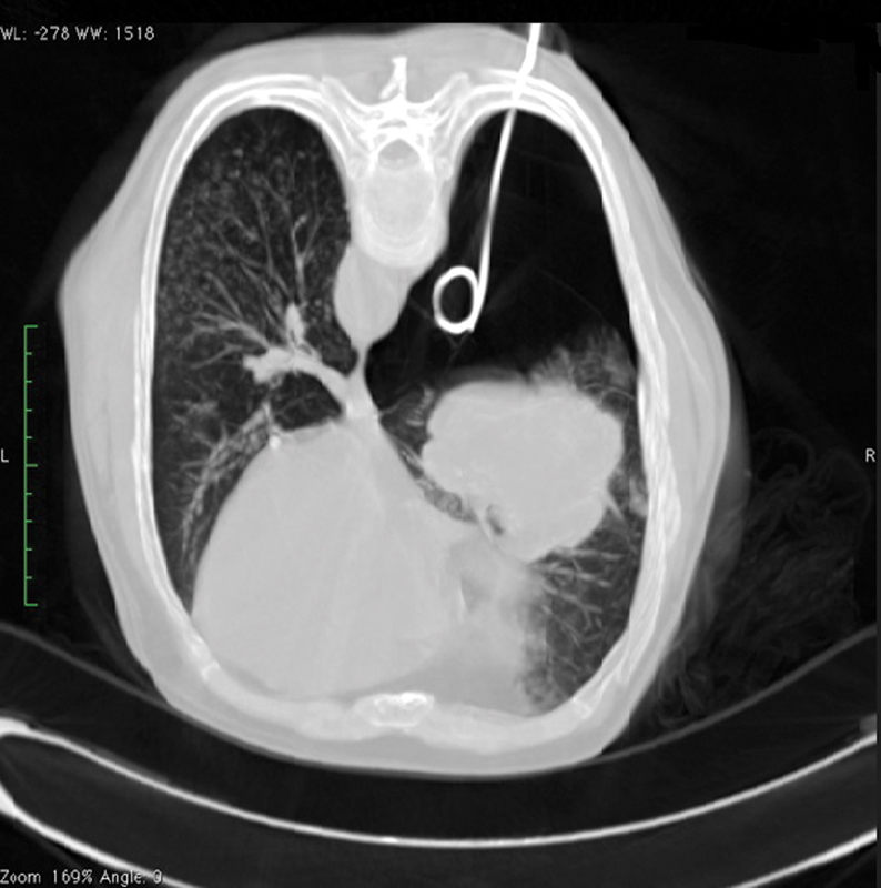
Of the 151 patients that developed pneumothorax, initially 91 (60.3%) had mild pneumothorax, 42 (27.8%) had moderate pneumothorax, and 18 (11.9%) had severe pneumothorax. However, on follow-up one case of mild pneumothorax and four cases of moderate pneumothorax progressed to severe pneumothorax. Therefore, in our study there were 90 (59.6%), 38 (25.2%), and 23 (15.2%) cases of mild, moderate, and severe pneumothorax, respectively ([Figs. 1],[2],[3]).


Initial Management
In mild pneumothorax cases only simple observation was done, in moderate pneumothoraces manual aspiration of pneumothorax was performed on table, and severe pneumothoraces were managed by 6F to 8F pigtail catheter insertion ([Fig. 4]).
Follow-Up: Check Radiograph
In the 91 patients of mild pneumothorax only 1 (1%) patient showed increase in size of pneumothorax on follow-up requiring catheter insertion. Rest of the 90 (99%) cases showed no or stable pneumothorax.
In the 42 cases of moderate pneumothorax who were managed by simple aspiration of pneumothorax, 4 (9.5%) cases showed increase in size of pneumothorax on follow-up requiring catheter insertion and 38 (90.5%) cases showed either no pneumothorax or only mild pneumothorax.
Final Management of Pneumothorax
Of the 151 cases of pneumothorax in our study 90 (59.6%) cases were successfully managed by simple observation, 38 (25.2%) cases were successfully managed by manual aspiration of pneumothorax, and 23 (15.2%) cases were successfully managed by catheter insertion. None of our patients required surgery.
Duration of Catheterization
A total 23 cases required pigtail catheter insertion in our study. These constituted 15.2% of pneumothorax cases and 6.4% of biopsy cases. The mean duration of catheterization in our study was 3.74 ± 1.09. The range was from 2 to 6 days ([Fig. 5]).

Discussion
CT-guided lung biopsy is usually performed in the outpatient setting to minimize costs and inconvenience to the patient. The problem most responsible for complicating outpatient management is not the presence of the pneumothorax per se, but the size of the pneumothorax with large, severe pneumothoraces requiring chest tube placement. Thus, efforts are required to decrease the necessity of chest tube placement for postbiopsy pneumothoraces. A variety of different techniques have been postulated to reduce the risk of worsening pneumothorax caused by biopsy and the need for chest tube placement. These include manual aspiration and maintaining a puncture site down position after biopsy, although the effect of positioning on reducing tube requirement is controversial.[3] [6] More aggressive methods to prevent worsening of pneumothorax include the placement of gel foam, glue, and autologous blood clots through the needle tract after biopsy; however, these have not yet found widespread acceptance.[7] In 1996, Yankelevitz et al reported success with manual aspiration at the time of biopsy for large pneumothoraces, thus avoiding chest tube placement in 70% of such cases.[8] This technique was further advocated by Yamagami et al in a series of studies in which they performed manual aspiration in all but mild pneumothoraces irrespective of symptoms.[6] [9] [10] [11] The mechanism of cure of the pneumothorax after manual aspiration is that the reapposition of the visceral and parietal pleural surfaces prevent leakage of air. We do not consider all patients to be good candidates for treatment with aspiration alone. In cases with severe pneumothorax or symptomatic pneumothorax the lung can recollapse during the observation period due to an enlarging pneumothorax and it might not be possible to immediately place a chest tube. Also, since all these cases have underlying lung lesions their respiratory reserve might be compromised. Hence, in the best interests of the patient, we prefer to place a catheter in cases with severe symptomatic pneumothoraces.
Our management strategy was predominantly determined by the size of the postprocedure pneumothorax whether mild, moderate, or severe. Of the 151 cases of pneumothorax in our study 90 cases were successfully managed by simple observation (59.6%), 38 (25.2%) cases were successfully managed by manual aspiration of pneumothorax, and 23 (15.2%) cases were successfully managed by catheter insertion. However, as we took a check CT prior to removal of needle out of lung, this might have resulted in underestimation of detection of pneumothorax in our study.
The majority of pneumothoraces (84.8%) therefore requires only observation or can be safely and effectively treated by manual aspiration on table without significantly increasing the morbidity of the patient, and thus required no overnight hospital stay and were discharged after few hours of observation. The overall catheter insertion rate was only 6.4% (15.2% of pneumothorax cases) and these were the only cases that increased the morbidity of the patient requiring hospitalization. The mean duration of catheterization in our study was 3.74 ± 1.09 days. The range was from 2 to 6 days. Chami et al report a chest tube placement rate of 15.3% (9/59 cases of pneumothorax) and rest of the cases were managed conservatively. Hiraki et al in their study report a chest tube placement rate of 11.9% (55/464) of the pneumothoraces. In the study conducted by Rizzo et al pneumothorax was treated by placing a chest drain in 13/72 cases (18% of patients with pneumothorax, 8% of all procedures).[12] [13] [14]
Yamagami et al evaluated the efficacy of simple aspiration of air from the pleural space to prevent increased pneumothorax and to avoid chest tube placement. They defined moderate or severe pneumothorax as a pneumothorax seen on more than seven CT slices (10 mm). Patients with a moderate or severe pneumothorax irrespective of the presence of symptoms underwent immediate manual aspiration while on the CT scanner table. Twenty of the 46 pneumothoraces (43.4%) were treated by manual aspiration, while 26 patients (56.5%) were simply observed. In 43 of the 46 pneumothoraces (93.5%), the pneumothorax resolved completely on follow-up chest radiographs without requiring tube placement. Only three patients (2.2% of the entire series; 6.5% of those who had pneumothorax) required chest tube placement. In our study the percentage of pneumothoraces that were simply observed and those requiring intervention was similar to their study (59.6% vs. 56.5% and 40.4% vs. 43.4%). However, our catheter insertion rate was higher than theirs (15.2% vs. 6.5%) because whereas we inserted catheters in all severe pneumothoraces they first performed aspiration in these cases.[9]
Yamagami et al subsequently reported a series of 102 postbiopsy pneumothorax cases. In 87 of these cases, pneumothorax disappeared spontaneously after manual aspiration. However, in the remaining 15 cases (14.7% of 102 cases), chest tube placement was finally required. In another series Yamagami et al had 243 pneumothorax cases, 112 (46.1%) of which were treated with manual aspiration immediately after biopsy. In 210 (86.4%) cases, the pneumothorax had resolved completely on follow-up chest radiographs without chest tube placement. Only 33(13.5%) patients required chest tube placement. Requirement of chest tube insertion significantly increased in parallel with the degree of pneumothorax as shown on postbiopsy CT images. The rate of chest tube insertion was statistically higher in subjects with values for aspirated air above 543 mL. In their study size of the pneumothorax (none, mild, moderate, severe) was the only variable associated with risk of chest tube insertion. These results are similar to our study. Note that 84.8% of our pneumothorax cases were managed without the need for catheter placement and only 15.2% of pneumothorax cases required catheterization.[6] [11]
Conclusion
-
Majority of pneumothoraces are benign and do not require any intervention, just observation.
-
Manual aspiration is an effective way of treating moderate pneumothoraces with success rate of 90%, thereby reducing the number of cases requiring catheter insertion. However, approximately 10% of these cases redevelop/progress to severe pneumothorax requiring pigtail insertion and therefore should be followed up at 2 to 3 hours.
-
A significant percentage of pneumothorax cases (15.2%) require catheter insertion resulting in increased morbidity of the patient requiring hospitalization. However, these are also safely and effectively managed with few days of pigtail catheterization.
Conflict of Interest
None declared.
Authors' Contributions
S.F.A. contributed with writing and concept design. S.T.A. contributed with data analysis and writing concept. S.O.A. took part in literature search and literature review. R.I. conducted literature search and literature review. C.N.A. contributed with editing, literature review, and preparation. G.T. contributed with data acquisition and data analysis. S.F. contributed with data analysis and data acquisition.
-
References
- 1 Haaga JR, Haaga TL, Wu H. Image Guided Interventions: CT emphasis. In: Haaga J, Dogra V, Forsting M, Gilkeson R, Ha H, Sundaram M. eds. CT and MRI of the Whole Body. 5th ed.. Philadelphia, USA: Mosby, Elsevier; 2009: 2462-2486
- 2 Wu CC, Maher MM, Shepard JA. Complications of CT-guided percutaneous needle biopsy of the chest: prevention and management. Am J Roentgenol 2011; 196 (06) W678-82
- 3 Moore EH, Shepard JA, McLoud TC, Templeton PA, Kosiuk JP. Positional precautions in needle aspiration lung biopsy. Radiology 1990; 175 (03) 733-735
- 4 Klose KC. CT-guided large-bore biopsy: extrapleural injection of saline for safe transpleural access to pulmonary lesions. Cardiovasc Intervent Radiol 1993; 16 (04) 259-261
- 5 Shepard JO. Complications of percutaneous needle aspiration biopsy of the chest: prevention and management. Semin Intervent Radiol 1994; 11: 181-186
- 6 Yamagami T, Kato T, Hirota T, Yoshimatsu R, Matsumoto T, Nishimura T. Usefulness and limitation of manual aspiration immediately after pneumothorax complicating interventional radiological procedures with the transthoracic approach. Cardiovasc Intervent Radiol 2006; 29 (06) 1027-1033
- 7 McCartney R, Tait D, Stilson M, Seidel GF. A technique for the prevention of pneumothorax in pulmonary aspiration biopsy. Am J Roentgenol Radium Ther Nucl Med 1974; 120 (04) 872-875
- 8 Yankelevitz DF, Davis SD, Henschke CI. Aspiration of a large pneumothorax resulting from transthoracic needle biopsy. Radiology 1996; 200 (03) 695-697
- 9 Yamagami T, Nakamura T, Iida S, Kato T, Nishimura T. Management of pneumothorax after percutaneous CT-guided lung biopsy. Chest 2002; 121 (04) 1159-1164
- 10 Yamagami T, Kato T, Iida S, Hirota T, Yoshimatsu R, Nishimura T. Efficacy of manual aspiration immediately after complicated pneumothorax in CT-guided lung biopsy. J Vasc Interv Radiol 2005; 16 (04) 477-483
- 11 Yamagami T, Terayama K, Yoshimatsu R, Matsumoto T, Miura H, Nishimura T. Role of manual aspiration in treating pneumothorax after computed tomography-guided lung biopsy. Acta Radiol 2009; 50 (10) 1126-1133
- 12 Chami HA, Faraj W, Yehia ZA. et al. Predictors of pneumothorax after CT-guided transthoracic needle lung biopsy: the role of quantitative CT. Clin Radiol 2015; 70 (12) 1382-1387
- 13 Hiraki T, Mimura H, Gobara H. et al. CT fluoroscopy-guided biopsy of 1,000 pulmonary lesions performed with 20-gauge coaxial cutting needles: diagnostic yield and risk factors for diagnostic failure. Chest 2009; 136 (06) 1612-1617
- 14 Rizzo S, Preda L, Raimondi S. et al. Risk factors for complications of CT-guided lung biopsies. Radiol Med (Torino) 2011; 116 (04) 548-563
Address for correspondence
Publication History
Article published online:
15 March 2023
© 2023. Indian Radiological Association. This is an open access article published by Thieme under the terms of the Creative Commons Attribution-NonDerivative-NonCommercial License, permitting copying and reproduction so long as the original work is given appropriate credit. Contents may not be used for commercial purposes, or adapted, remixed, transformed or built upon. (https://creativecommons.org/licenses/by-nc-nd/4.0/)
Thieme Medical and Scientific Publishers Pvt. Ltd.
A-12, 2nd Floor, Sector 2, Noida-201301 UP, India
-
References
- 1 Haaga JR, Haaga TL, Wu H. Image Guided Interventions: CT emphasis. In: Haaga J, Dogra V, Forsting M, Gilkeson R, Ha H, Sundaram M. eds. CT and MRI of the Whole Body. 5th ed.. Philadelphia, USA: Mosby, Elsevier; 2009: 2462-2486
- 2 Wu CC, Maher MM, Shepard JA. Complications of CT-guided percutaneous needle biopsy of the chest: prevention and management. Am J Roentgenol 2011; 196 (06) W678-82
- 3 Moore EH, Shepard JA, McLoud TC, Templeton PA, Kosiuk JP. Positional precautions in needle aspiration lung biopsy. Radiology 1990; 175 (03) 733-735
- 4 Klose KC. CT-guided large-bore biopsy: extrapleural injection of saline for safe transpleural access to pulmonary lesions. Cardiovasc Intervent Radiol 1993; 16 (04) 259-261
- 5 Shepard JO. Complications of percutaneous needle aspiration biopsy of the chest: prevention and management. Semin Intervent Radiol 1994; 11: 181-186
- 6 Yamagami T, Kato T, Hirota T, Yoshimatsu R, Matsumoto T, Nishimura T. Usefulness and limitation of manual aspiration immediately after pneumothorax complicating interventional radiological procedures with the transthoracic approach. Cardiovasc Intervent Radiol 2006; 29 (06) 1027-1033
- 7 McCartney R, Tait D, Stilson M, Seidel GF. A technique for the prevention of pneumothorax in pulmonary aspiration biopsy. Am J Roentgenol Radium Ther Nucl Med 1974; 120 (04) 872-875
- 8 Yankelevitz DF, Davis SD, Henschke CI. Aspiration of a large pneumothorax resulting from transthoracic needle biopsy. Radiology 1996; 200 (03) 695-697
- 9 Yamagami T, Nakamura T, Iida S, Kato T, Nishimura T. Management of pneumothorax after percutaneous CT-guided lung biopsy. Chest 2002; 121 (04) 1159-1164
- 10 Yamagami T, Kato T, Iida S, Hirota T, Yoshimatsu R, Nishimura T. Efficacy of manual aspiration immediately after complicated pneumothorax in CT-guided lung biopsy. J Vasc Interv Radiol 2005; 16 (04) 477-483
- 11 Yamagami T, Terayama K, Yoshimatsu R, Matsumoto T, Miura H, Nishimura T. Role of manual aspiration in treating pneumothorax after computed tomography-guided lung biopsy. Acta Radiol 2009; 50 (10) 1126-1133
- 12 Chami HA, Faraj W, Yehia ZA. et al. Predictors of pneumothorax after CT-guided transthoracic needle lung biopsy: the role of quantitative CT. Clin Radiol 2015; 70 (12) 1382-1387
- 13 Hiraki T, Mimura H, Gobara H. et al. CT fluoroscopy-guided biopsy of 1,000 pulmonary lesions performed with 20-gauge coaxial cutting needles: diagnostic yield and risk factors for diagnostic failure. Chest 2009; 136 (06) 1612-1617
- 14 Rizzo S, Preda L, Raimondi S. et al. Risk factors for complications of CT-guided lung biopsies. Radiol Med (Torino) 2011; 116 (04) 548-563