
RSS-Feed abonnieren
DOI: 10.1055/s-0042-1757429
Radiological and Morphometric Study of the Emissary Foramens and Canal in the Posterior Cranial Fossa of the Human Skull with Its Neurosurgical Significance
Abstract
Objective The posterior condylar canals (PCCs), posterior condylar veins (PCVs), occipital foramen (OF), and occipital emissary vein (OEV) are potential anatomical landmarks for surgical approaches through the lateral foramen magnum. We performed the study to make morphometric and radiological analyses of the various emissary foramens and vein in the posterior cranial fossa.
Methods Morphometric study were performed on 95 dry occipital bones and radiological analyses on computed tomography (CT) angiography images of 150 patients. The number of OFs on both sides was recorded and PCC length and mean diameters of the internal and external orifices of PCC were measured for bony specimens. Prevalence of PCV and PCV size was investigated using CT angiography.
Results Mean PCC length was higher in the left side (9.85 ± 2.5). Mean diameter of the internal orifice and the external orifice diameter were almost the same. The majority of PCCs (75–79.33%) had 2 to 5 mm diameter; only 4 to 9.2% were small in size (< 2 mm). In CT angiography, PCV was not identified in 23 (15.33%) patients. PCVs were located bilaterally in 105 (70%) and unilaterally in 22 (20.5%) patients. Only 11.3% of PCVs were large in size (> 5 mm), 80% of PCVs were medium sized (2–5 mm), and 8.6% were small sized (< 2 mm).
Conclusion Normal values of OF, PCC, PCV, and OEV could serve as a future reference for the understanding of the physiology of craniocervical venous drainage, which is necessary to avoid surgical complications and can also serve as a guide to surgical interventions for pathologies of the posterior cranial fossa, such as tumors and injuries.
#
Keywords
occipital foramen - angiography - computed tomography - emissary vein - posterior condylar canal - posterior condylar veinIntroduction
Emissary veins are valveless veins that pass through cranial foramina. Along with the internal jugular vein, they connect the extracranial venous system with intracranial venous sinuses.[1] Posterior condylar foramen (PCF), the foramen that posterior condylar vein (PCV) passes through, is the largest emissary foramen in the retromastoid region.[2] [3] [4] Occipital emissary vein (OEV) passes through the occipital canal (OC) and provides venous drainage from the transverse sinus to the vertebral venous plexus via the occipital vein. Altogether PCV, OEV, and mastoid emissary vein (MEV) are the major posterior fossa emissary veins. It is important to recognize and describe these veins to prevent complications during surgeries.[5]
These veins protect the brain from increases in intracranial pressure in patients with lesions of the neck or skull base and obstructed internal jugular veins[6]; they also play an important role in cooling venous blood circulating through cephalic structures.[7] In several instances, emissary veins may be enlarged in patients with high-flow vascular malformations or severe hypoplasia or aplasia of the jugular veins. They are associated with craniofacial syndromes.[2] [8] Dilated emissary veins may cause tinnitus.[3] Also, these valveless veins connecting the extracranial venous system with intracranial venous sinuses may transfer infections from the superficial tissues into the cranium.[9]
Inadvertent injury of these vessels poses a significant problem as such air embolism or thromboembolism and recognition of these structures and releasing it from other surrounding tissues are required to protect the veins from avulsion or laceration that cause alarming hemorrhage.[10] In the literature survey, there were very few studies available stating the importance of emissary foramina and veins and there was no study conducted in Indian population till date. Therefore, a detailed study and knowledge about the PCC, PCV, OC, and OEV are needed. This study was performed to make morphometric and radiological analyses of the occipital foramen (OF), PCC, PCV, and OEV in Indian population.
#
Materials and Methods
Dry Bones
This study was performed on 100 human adult dry occipital bones (Indian ethnicity) obtained from the collections of the anatomy laboratory in our institution. Five occipital bones were excluded due to damaged condylar canals. Morphometric measurements such as PCC length and mean diameters of the internal and external orifices of PCC were measured by taking the average of transverse and sagittal diameter using a standard stainless caliper (Mitutoyo 532–101, Tokyo, Japan). The number of OF on both sides was recorded. All measurements were performed by the same researcher.
#
Radiological Images
The radiological part of this study was performed at Department of Radiodiagnosis, Advanced Medical Research Institute (AMRI) Hospital Ltd, Kolkata, India. The study was conducted according to the Helsinki Declaration. Ethical approval was obtained from the institute ethical committee (Number-EC/2018/10). Computed tomography (CT) angiography images of 175 patients referred to the hospital for investigation of vascular pathologies were reviewed. Patients were scanned with a 64-slice, positron emission tomography/computed tomography (Discovery PET/CT 690, GE Healthcare, United States) with the following settings: beam collimation, 160 × 0.5 mm; rotation time, 0.5s; pitch, 0.869; section thickness and intervals, 0.5 mm and 0.4 mm; image matrix, 512 × 512; and field of view, 240 mm. The tube current at 120 kVp was in the range of 100 to 500 mA. Bolus-tracking and monitor scan was used to monitor changes in region of interest (ROI) intensity and trigger the scanning when a threshold was reached. ROI for bolus tracking was fixed at carotid artery. A 60-mL bolus of nonionic contrast media (Omnipaque, Iodine 300 mgI/mL; GE Healthcare, Chicago, Illinois, United States) was administered into an antecubital vein by using a pressure injector (MEDRAD Stellant CT Injection, Bayers AG, United States) with an injection rate of 5 mL/s. Multidetector CT angiography data were transferred from the archive to a work station (ADW workstation, GE Healthcare) providing three-dimensional postprocessing options, multiplanar image reformatting, and maximum intensity projections. Out of 175 patients, 18 patients were excluded from the study for inadequate image quality mainly due to patient movement and technical problems, and seven patients were excluded because they had mastoid region and/or posterior fossa surgery prior to CT angiography. The mean age of the 150 patients (62 females, 88 males) was 55.4 years. The prevalence of the PCV was recorded for both sides as absent, unilateral, or bilateral. PCV was classified by size into three groups: small (< 2 mm), medium-sized (2–5 mm), and large (> 5 mm).
#
Statistical Analysis
The data were analyzed using the SPSS version 20 (SPSS Inc., Chicago, Illinois, United States). Descriptive statistics with means and standard deviations were computed for each measurement in both sexes. Student's t-test was used to compare the means of right and left sides for PCC. p-Value less than 0.05 was accepted as statistically significant.
#
#
Results
Dry Bones
It was observed that the 5 out of the 95 occipital bones had OFs in the midline (5.26%) ([Fig. 1A]), 75 occipital bones were bilateral (78.9%) ([Fig. 1B]), and 10 were unilateral (10.5%) ([Fig. 1C]). The foramen was absent bilaterally in three bones (3.7%) ([Fig. 1D]). In two occipital bone (2.1%), bilateral OFs located near the midline formed canal ([Fig. 1E]) ([Table 1]). PCC was located in the left side of 65 bones (68.42%) out of the 95 occipital bones, while 60 bones (63.15%) had their PCC on the right side. Mean length of the PCC was measured 9.85 ± 2.5 (17–1 mm) and 8.75 ± 2.5 (18–2 mm) on the left and right sides, respectively. The mean diameter of the internal orifice was 3.20 ± 1.37 mm (13–1 mm) on the left and 3.0 ± 2.42 mm (10–1 mm) on the right side. For the external orifice, the mean diameter was measured 3.90 ± 1.54 mm (10–1 mm) on the left and 3.55 ± 3.1 mm (9–1 mm) on the right side ([Table 2]). No significant differences were found in the mean length and in the diameters of the internal and external orifices between the right and left sides (p > 0.05) ([Table 2]). Morphometric measurement showed that majority of PCCs (75–78%) of occipital bone had the diameter between 2 and 5 mm; only 3 to 9% were small in size (< 2 mm) ([Table 3]).
Abbreviations: PCC, posterior condylar canal; SD, standard deviation.
Abbreviation: PCC, posterior condylar canal.

#
Radiological Images
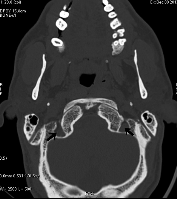
In CT angiography images, PCV was not identified in 23 (15.33%) of the 150 patients. PCVs were located bilaterally in 105 (70%) and unilaterally in 22 (14.66%) patients. The number of unilateral right-sided PCVs (n = 12) was slightly higher than the unilateral left-sided PCVs (n = 10) ([Table 4]). The majority of PCVs were medium-sized (diameter 2–5 mm) found in 80% (n = 120), and 8.6% (n = 13) were small-sized (< 2 mm) ([Figs. 2] [3] [4] [5]) and only 11.3% of PCVs (n = 17) were large in diameter (> 5 mm) ([Table 5]).


Abbreviations: CT, computed tomography; PCV, posterior condylar vein.
|
Diameter |
Prevalence, n (%) |
|---|---|
|
< 2 mm |
13 (8.6) |
|
2–5 mm |
120 (80) |
|
> 5 mm |
17 (11.3) |
Abbreviations: CT, computed tomography; PCV, posterior condylar vein.
#
#
Discussion
Morphological and radiological studies on OF, PCC, PCV, OEV, and other EVs have become a crucial part of diagnosis in neurosurgery,[9] but the absence of important anatomic data on these foramina is a severe deficiency in available literature. The occipital emissary foramen and PCFs are present in the squamous part of the occipital bone at the occipital protuberance and transmits a vein that connects the confluence of the dural venous sinuses with veins of the suboccipital venous plexus.[10] The morphological features of PCFs are significant specifically for neurosurgical procedures in posterior cranial fossa.[11]
Following different courses of PCC and PCV, and there exists several types of intracranial orifice connections of PCV in the cranial base. Besides, the shift in location of the intracranial orifice indicate the presence of other venous structures, such as marginal sinus or occipital sinus, which rarely leave any visible routes on occipital bones.[12]
The posterior condylar canal forms a communication between the jugular foramen and the condylar fossa just posterior to the occipital condyles.[11] It transmits an emissary vein that allows anastomosis of the jugular bulb or sigmoid sinus to the suboccipital venous plexus.[12] [13] The patient with prominent posterior condylar canal may have some clinical relevance resulting in venous anastomoses that may allow for alternative venous drainage and, therefore, the theoretical possibility of a false-positive Queckenstedt's test, suggesting spinal block.[8] While CT angiography is a successful technique for imaging vascular structures, it does not provide reliable results for the imaging of bone structures. Additionally, the discrepancy between radiological and morphological data in this study may be because an enlarged emissary foramen does not necessarily transmit a large EV.[13]
In a previous study by Matsushima et al[12] on two dry skulls and Özgören et al[14] on 91 occipital bones measured the internal orifice diameter of PCC as 3.5 and 3.87 mm, external orifice diameter as 3.95 mm, and mean PCC as length 6.8 and 10.31 mm. In our study, these finding were similar to the previous studies, except for the mean length of PCC that was slightly higher in our study which concurs with the study by Özgören et al,[14] which measured as 9.85 (17–1) mm on the left and 8.75 (18–2) mm on the right side. An abnormal posterior condylar emissary vein can be associated with clinical presentation objective pulsatile tinnitus. Lambert and Cantrell et al[4] and Lee et al[6] investigated the relation between objective tinnitus and EV flow and reported a case of objective tinnitus associated with an abnormal PCV and MEV using Doppler ultrasound and imaging. Forte et al[15] by Doppler method studied posterior auricular region on 30 asymptomatic human subjects and observed PCV in 84%; none was found to emissary vein flow. The prevalence of PCV found in this study was 84.66%. Recognition of the normal vein of the posterior condylar canal may prevent misinterpretation of the vein as a neoplasm or an abnormal lymph node at CT or magnetic resonance imaging (MRI).[15]
The incidence of PCC in various population reported by several studies can be seen in literature. In a first ever study by Boyd,[16] presence of PCC was reported in both sides in 46.6% of skulls, the higher percentage than in the case of any other foramen. Boyd also found that PCC is absent on both sides in only 23.1% and he also noted that unilateral condylar foramen is present rather more often on right side than on the left side (16.5 and 13.8%). In Australian skulls, PCC is smaller than in other races and is less often present in the proportion of 67:77. The condyloid foramen is the largest and most constant (77%) of all the foramina in man. A study by Falk[17] reported the incidence of posterior condylar canal to 60% in Australopithecines race. Wysocki et al[18] conducted the study on 100 macerated human skull of polish origin and reported the presence of PCC to 65%. Krause[19] found 21% bilaterally presence, while unilateral was 38% of skulls. Another study reported posterior condylar canals were identified in 36 of 50 sides in dry bones [12] Similarly, a study by Natsis et al[20] reported incidence of PCF is 75.5% of 143 Greek adult dry skulls and absent in 33 sides (33%). Ginsberg[21] in his study identified condylar canal unilaterally in 50% of the cases and bilaterally in 30%. Similarly on Turkish population, a study has reported that PCC was absent unilaterally in 27% of specimens and bilaterally in 17% of the skulls and he concluded that posterior condylar canal may not be used as a constant landmark.[22]
Various studies conducted on dry skull of Indian origin have reported that posterior condylar canal was present in 9.7% out of which 6.0% were on the left side and 3.6% bilateral in position[23] and similarly study on south Indian population was noted in which PCC incidence was 16%.[24] Contrary to the previous results, another study reported the higher incidence of PCC to 78.9% and mentioned that the PCC was doubled in six of the 144 patent foramina (4%) and tripled in one case.[25] Similar to the previous results in this study, the overall incidence of PCC was 70% ([Table 6]) with unilateral 10.59% and bilateral 78.9% and PCC is a reliable and most constant foramina in man.
|
Authors/year |
Population |
Overall incidence of PCC (%) |
|---|---|---|
|
Boyd 1930[16] |
Australian and New Zealand |
75.5 |
|
Falk 1986[17] |
Australopithecines |
60.0 |
|
Wysocki et al 2006[18] |
Polish |
65.0 |
|
Krause 1988[19] |
Spain |
21.0 |
|
Natsis et al 2013[20] |
Greek |
75.5 |
|
Ginsberg 1994[21] |
American |
55.9 |
|
Avci et al 2011[22] |
Turkish |
66.0 |
|
Chauhan and Sharma 2013[23] |
North Indian |
3.6 |
|
Kothandaraman and Lokanadham 2015[24] |
Indian |
16.0 |
|
Kavitha and Anand 2013[25] |
South Indian |
78.9 |
|
Present study |
South Indian |
70.0 |
Knowledge of anatomy of the cranial emissary foramens and veins provides some advantages during endovascular treatments and posterior fossa surgeries. The posterior condylar canal and other emissary channels may allow for the venous escape of blood in the event of unilateral or bilateral jugular venous obstruction acting as an alternative route of venous drainage from the brain during surgeries. It is possible that in the event of a high-flow state or vascular malformation, the PCC could become enlarged.[26]
#
Conclusion
The results of our study contribute to the research on posterior cranial foramina mainly posterior condylar canal and provide familiarity with the anatomy and the basic variations in the venous network at the posterior cranial fossa essential for accurate radiologic diagnosis and can prevent misinterpretation of the normal findings. It should not be misinterpreted as abnormal such as tumors, condylar fractures, and other injuries. With the increased advent of application of MRI and CT, the foramina of the skull are being observed proficiently in the clinical setup than earlier.
#
#
Conflict of interest
None declared.
Ethical Approval and Informed Consent
The study is approved by Institutional Ethics committee (Number-EC/2018/10), Kannur Hospital, Anjarakandy, Kerala, India. Consent has been taken by study participants and the study is conducted in accordance with Helsinki declaration.
Authors' Contributions
M.A.A. and T.M.H.G. were involved in protocol/project development. A.N., D.M., and T.M.H.G. collected the data. M.A.A. and T.M.H.G. analyzed the data and contributed to manuscript writing/editing. All authors have read and approved the final version of the manuscript.
-
References
- 1 Mortazavi MM, Tubbs RS, Riech S. et al. Anatomy and pathology of the cranial emissary veins: a review with surgical implications. Neurosurgery 2012; 70 (05) 1312-1318 , discussion 1318–1319
- 2 Coin CG, Malkasian DR. (1971). Foramen magnum. In: Newton TH, Potts DG, eds. Radiology of the Skull and Brain: The Skull. St. Louis: Mosby; 275-347
- 3 Jeevan DS, Anlsow P, Jayamohan J. Abnormal venous drainage in syndromic craniosynostosis and the role of CT venography. Childs Nerv Syst 2008; 24 (12) 1413-1420
- 4 Lambert PR, Cantrell RW. Objective tinnitus in association with an abnormal posterior condylar emissary vein. Am J Otol 1986; 7 (03) 204-207
- 5 Lanzieri CF, Duchesneau PM, Rosenbloom SA, Smith AS, Rosenbaum AE. The significance of asymmetry of the foramen of Vesalius. AJNR Am J Neuroradiol 1988; 9 (06) 1201-1204
- 6 Lee S-H, Kim SS, Sung K-Y, Nam EC. Pulsatile tinnitus caused by a dilated mastoid emissary vein. J Korean Med Sci 2013; 28 (04) 628-630
- 7 Pekcevik Y, Sahin H, Pekcevik R. Prevalence of clinically important posterior fossa emissary veins on CT angiography. J Neurosci Rural Pract 2014; 5 (02) 135-138
- 8 Pekçevik Y, Pekçevik R. Why should we report posterior fossa emissary veins?. Diagn Interv Radiol 2014; 20 (01) 78-81
- 9 Louis Jr RG, Loukas M, Wartmann CT. et al. Clinical anatomy of the mastoid and occipital emissary veins in a large series. Surg Radiol Anat 2009; 31 (02) 139-144
- 10 Marsot-Dupuch K, Gayet-Delacroix M, Elmaleh-Bergès M, Bonneville F, Lasjaunias P. The petrosquamosal sinus: CT and MR findings of a rare emissary vein. AJNR Am J Neuroradiol 2001; 22 (06) 1186-1193
- 11 Wilson MH, Newman S, Imray CH. The cerebral effects of ascent to high altitudes. Lancet Neurol 2009; 8 (02) 175-191
- 12 Matsushima K, Kawashima M, Matsushima T, Hiraishi T, Noguchi T, Kuraoka A. Posterior condylar canals and posterior condylar emissary veins-a microsurgical and CT anatomical study. Neurosurg Rev 2014; 37 (01) 115-126
- 13 Tanoue S, Kiyosue H, Sagara Y. et al. Venous structures at the craniocervical junction: anatomical variations evaluated by multidetector row CT. Br J Radiol 2010; 83 (994) 831-840
- 14 Ozgören O, Güleç F, Pekçevik Y, Fengül G. Radiological and anatomical evaluation of the posterior condylar canal, posterior condylar vein and occipital foramen. Anatomy 2015; 9 (03) 151-155
- 15 Forte V, Turner A, Liu P. Objective tinnitus associated with abnormal mastoid emissary vein. J Otolaryngol 1989; 18: 232-235
- 16 Boyd GI. The emissary foramina of the cranium in man and the anthropoids. J Anat 1930; 65 (Pt 1): 108-121
- 17 Falk D. Evolution of cranial blood drainage in hominids: enlarged occipital/marginal sinuses and emissary foramina. Am J Phys Anthropol 1986; 70 (03) 311-324
- 18 Wysocki J, Reymond J, Skarzyński H, Wróbel B. The size of selected human skull foramina in relation to skull capacity. Folia Morphol (Warsz) 2006; 65 (04) 301-308
- 19 Krause W. The posterior condylar canal. In: Testut L and Latarjet A, eds. Treaty of Human Anatomy. Barcelona: Salvat; 1988. 1. 152-158
- 20 Natsis K, Piagkou M, Skotsimara G, Piagkos G, Skandalakis P. A morphometric anatomical and comparative study of the foramen magnum region in a Greek population. Surg Radiol Anat 2013; 35 (10) 925-934
- 21 Ginsberg LE. The posterior condylar canal. AJNR Am J Neuroradiol 1994; 15 (05) 969-972
- 22 Avci E, Dagtekin A, Ozturk AH. et al. Anatomical variations of the foramen magnum, occipital condyle and jugular tubercle. Turk Neurosurg 2011; 21 (02) 181-190
- 23 Chauhan K, Sharma A. Osteotic variations in paracondylar region of adult human skulls: Incidence and clinical significance. Int J Anat Res 2013; 03: 158-160
- 24 Kothandaraman U, Lokanadham S. Posterior condylar foramen—anatomical variation. Int J Med Sci Public Health 2015; 4 (02) 222-224
- 25 Kavitha S, Anand A. A study of the condylar canal and its incidence, morphology and clinical significance. Int J Curr Res Rev 2013; 5 (02) 66-70
- 26 Weissman JL. Condylar canal vein: unfamiliar normal structure as seen at CT and MR imaging. Radiology 1994; 190 (01) 81-84
Address for correspondence
Publikationsverlauf
Artikel online veröffentlicht:
28. Oktober 2022
© 2022. Asian Congress of Neurological Surgeons. This is an open access article published by Thieme under the terms of the Creative Commons Attribution-NonDerivative-NonCommercial License, permitting copying and reproduction so long as the original work is given appropriate credit. Contents may not be used for commercial purposes, or adapted, remixed, transformed or built upon. (https://creativecommons.org/licenses/by-nc-nd/4.0/)
Thieme Medical and Scientific Publishers Pvt. Ltd.
A-12, 2nd Floor, Sector 2, Noida-201301 UP, India
-
References
- 1 Mortazavi MM, Tubbs RS, Riech S. et al. Anatomy and pathology of the cranial emissary veins: a review with surgical implications. Neurosurgery 2012; 70 (05) 1312-1318 , discussion 1318–1319
- 2 Coin CG, Malkasian DR. (1971). Foramen magnum. In: Newton TH, Potts DG, eds. Radiology of the Skull and Brain: The Skull. St. Louis: Mosby; 275-347
- 3 Jeevan DS, Anlsow P, Jayamohan J. Abnormal venous drainage in syndromic craniosynostosis and the role of CT venography. Childs Nerv Syst 2008; 24 (12) 1413-1420
- 4 Lambert PR, Cantrell RW. Objective tinnitus in association with an abnormal posterior condylar emissary vein. Am J Otol 1986; 7 (03) 204-207
- 5 Lanzieri CF, Duchesneau PM, Rosenbloom SA, Smith AS, Rosenbaum AE. The significance of asymmetry of the foramen of Vesalius. AJNR Am J Neuroradiol 1988; 9 (06) 1201-1204
- 6 Lee S-H, Kim SS, Sung K-Y, Nam EC. Pulsatile tinnitus caused by a dilated mastoid emissary vein. J Korean Med Sci 2013; 28 (04) 628-630
- 7 Pekcevik Y, Sahin H, Pekcevik R. Prevalence of clinically important posterior fossa emissary veins on CT angiography. J Neurosci Rural Pract 2014; 5 (02) 135-138
- 8 Pekçevik Y, Pekçevik R. Why should we report posterior fossa emissary veins?. Diagn Interv Radiol 2014; 20 (01) 78-81
- 9 Louis Jr RG, Loukas M, Wartmann CT. et al. Clinical anatomy of the mastoid and occipital emissary veins in a large series. Surg Radiol Anat 2009; 31 (02) 139-144
- 10 Marsot-Dupuch K, Gayet-Delacroix M, Elmaleh-Bergès M, Bonneville F, Lasjaunias P. The petrosquamosal sinus: CT and MR findings of a rare emissary vein. AJNR Am J Neuroradiol 2001; 22 (06) 1186-1193
- 11 Wilson MH, Newman S, Imray CH. The cerebral effects of ascent to high altitudes. Lancet Neurol 2009; 8 (02) 175-191
- 12 Matsushima K, Kawashima M, Matsushima T, Hiraishi T, Noguchi T, Kuraoka A. Posterior condylar canals and posterior condylar emissary veins-a microsurgical and CT anatomical study. Neurosurg Rev 2014; 37 (01) 115-126
- 13 Tanoue S, Kiyosue H, Sagara Y. et al. Venous structures at the craniocervical junction: anatomical variations evaluated by multidetector row CT. Br J Radiol 2010; 83 (994) 831-840
- 14 Ozgören O, Güleç F, Pekçevik Y, Fengül G. Radiological and anatomical evaluation of the posterior condylar canal, posterior condylar vein and occipital foramen. Anatomy 2015; 9 (03) 151-155
- 15 Forte V, Turner A, Liu P. Objective tinnitus associated with abnormal mastoid emissary vein. J Otolaryngol 1989; 18: 232-235
- 16 Boyd GI. The emissary foramina of the cranium in man and the anthropoids. J Anat 1930; 65 (Pt 1): 108-121
- 17 Falk D. Evolution of cranial blood drainage in hominids: enlarged occipital/marginal sinuses and emissary foramina. Am J Phys Anthropol 1986; 70 (03) 311-324
- 18 Wysocki J, Reymond J, Skarzyński H, Wróbel B. The size of selected human skull foramina in relation to skull capacity. Folia Morphol (Warsz) 2006; 65 (04) 301-308
- 19 Krause W. The posterior condylar canal. In: Testut L and Latarjet A, eds. Treaty of Human Anatomy. Barcelona: Salvat; 1988. 1. 152-158
- 20 Natsis K, Piagkou M, Skotsimara G, Piagkos G, Skandalakis P. A morphometric anatomical and comparative study of the foramen magnum region in a Greek population. Surg Radiol Anat 2013; 35 (10) 925-934
- 21 Ginsberg LE. The posterior condylar canal. AJNR Am J Neuroradiol 1994; 15 (05) 969-972
- 22 Avci E, Dagtekin A, Ozturk AH. et al. Anatomical variations of the foramen magnum, occipital condyle and jugular tubercle. Turk Neurosurg 2011; 21 (02) 181-190
- 23 Chauhan K, Sharma A. Osteotic variations in paracondylar region of adult human skulls: Incidence and clinical significance. Int J Anat Res 2013; 03: 158-160
- 24 Kothandaraman U, Lokanadham S. Posterior condylar foramen—anatomical variation. Int J Med Sci Public Health 2015; 4 (02) 222-224
- 25 Kavitha S, Anand A. A study of the condylar canal and its incidence, morphology and clinical significance. Int J Curr Res Rev 2013; 5 (02) 66-70
- 26 Weissman JL. Condylar canal vein: unfamiliar normal structure as seen at CT and MR imaging. Radiology 1994; 190 (01) 81-84