
RSS-Feed abonnieren
DOI: 10.1055/s-0042-1751036
Lipid-Based Nanocarrier Systems for Drug Delivery: Advances and Applications

- Introduction
- Traditional Lipid-Based Nanocarriers
- Lipid-Based Nanoemulsions
- Lipid-Based Nanomicelles
- Stimuli-Responsive Lipid-Based Nanocarriers
- Ionizable Lipid Nanoparticles
- Lipid–Polymer Nanocarriers
- Biomembrane-Camouflaged Nanocarriers
- Solid Lipid Nanoparticle and Nanostructured Lipid Carriers
- Lipid–Inorganic Nanocarriers
- Current Status and Future Perspectives
- Clinical Translation
- Industrial Production
- Future Perspectives
- Conclusion
- References
Abstract
Lipid-based nanocarriers have been extensively investigated for drug delivery due to their advantages including biodegradability, biocompatibility, nontoxicity, and nonimmunogenicity. However, the shortcomings of traditional lipid-based nanocarriers such as insufficient targeting, capture by the reticuloendothelial system, and fast elimination limit the efficiency of drug delivery and therapeutic efficacy. Therefore, a series of multifunctional lipid-based nanocarriers have been developed to enhance the accumulation of drugs in the lesion site, aiming for improved diagnosis and treatment of various diseases. In this review, we summarized the advances and applications of lipid-based nanocarriers from traditional to novel functional lipid preparations, including liposomes, stimuli-responsive lipid-based nanocarriers, ionizable lipid nanoparticles, lipid hybrid nanocarriers, as well as biomembrane-camouflaged nanoparticles, and further discussed the challenges and prospects of this system. This exploration may give a complete idea viewing the lipid-based nanocarriers as a promising choice for drug delivery system, and fuel the advancement of pharmaceutical products by materials innovation and nanotechnology.
#
Keywords
lipid-based nanocarriers - drug delivery - nanomedicine - lipid materials - clinical applicationIntroduction
The conventional “free” drugs in the clinical application always exhibit some common problems, such as poor solubility, unfavorable pharmacokinetics, suboptimal biodistribution, and lack of selectivity for target tissues.[1] A drug delivery system (DDS) possesses many advantages over free drug molecules, showing enhanced solubility and improved pharmacokinetics and therapeutic efficacy.[1] Due to the nano size and shape, a nanodrug delivery system could achieve theoretically superior drug delivery efficiency through specific tissue targeting and enhanced cell internalization and transportation to specific organelles.[2] [3] [4] However, the toxicology of nanocarriers always reminds an important issue, especially for medical use.[5] [6] It has been reported that polymeric and metallic nanocarriers may be toxic to the lung, kidney, or central nervous system.[7] Therefore, scientists have shown great interest in lipid-based nanocarriers with the advantages of being biodegradable, biocompatible, nontoxic, and nonimmunogenic. Lipid-based nanocarriers show great potential in augmenting bioavailability, improving pharmacokinetic profile, reducing toxicity, and achieving the desired concentration of the drug at otherwise inaccessible sites through various mechanisms.[8]
As the first liposome preparation, Doxil approved by Food and Drug Administration (FDA) in 1995 opened a new era of drug delivery using lipid-based nanocarriers. With in-depth research, lipid-based nanocarriers have achieved good results in drug delivery. The initial liposomes were only composed of phospholipids and some pharmaceutical excipients, which led to poor stability and accumulation both in vivo and in vitro. Researchers later found that some modifications on the surface of liposomes could provide steric hindrance and improve the stability of liposomes, and the most classic modification is PEGylation.[9] Moreover, other shortcomings of liposomes have been found, including insufficient targeting, rapid elimination from the blood, and capture by the reticuloendothelial system.[10] In recent years, a series of multifunctional lipid-based nanocarriers have been developed to improve the accumulation of drugs in the lesion site for the diagnosis and treatment of various diseases. The rational design of the modification on lipid-based nanocarriers was explored, including proteins, small molecular compounds, and even some synthetic polymer materials, to prolong the half-life of the nanocarriers in vivo and cross various pathological barriers for lesion-site targeting. With more in-depth understanding of diseases, drugs delivered by lipid-based nanocarriers are not only small molecular drugs, but also biomacromolecules such as antibodies, messenger RNA (mRNA), and small-interfering RNA (siRNA). At the same time, a large number of experiments are performed to verify and optimize the lipid-based nanocarriers to meet various requirements of drug delivery.[11] [12] [13] [14]
Lipid-based nanocarriers demonstrate advantages including improved bioavailability, enhanced tumor targeting, and reduced toxicity, showing great potential in clinical translation. In this review, we briefly summarized the characteristics and applications of some traditional lipid preparations, and emphatically introduced the advanced lipid-based nanocarriers. In addition, we further discuss prospects and challenges of the lipid-based nanocarriers.
#
Traditional Lipid-Based Nanocarriers
Liposomes
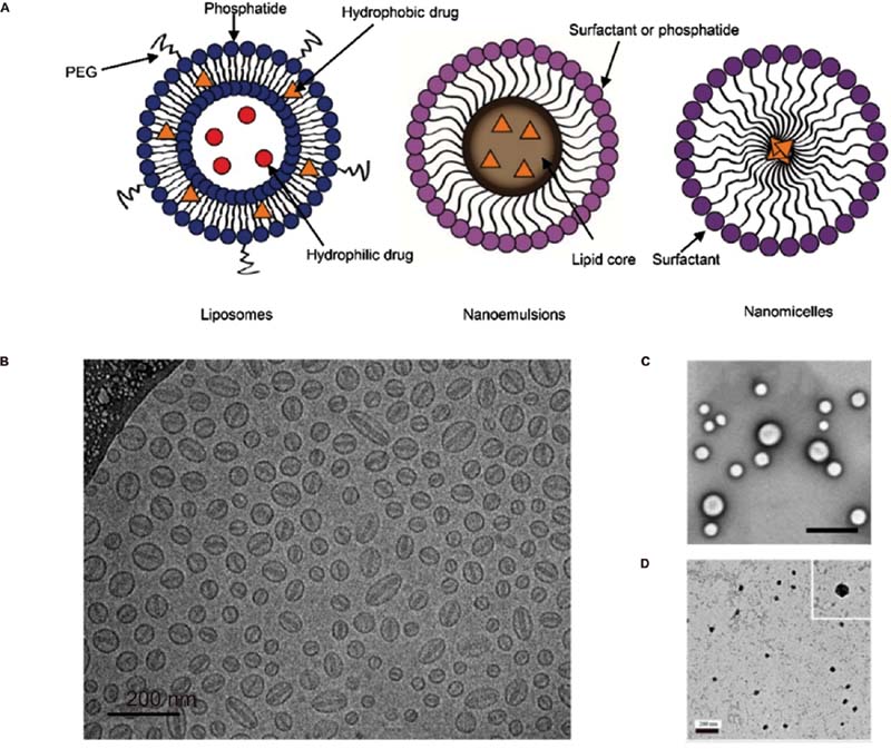
Since liposomes were first discovered by Bangham in the early 1960s,[15] and they have been widely studied and applied in drug delivery due to their unique lipid bilayer structure and characteristics ([Fig. 1]). Up to now, many liposome preparations have been approved for treatment of bacterial and fungal infections, cancer, and other diseases ([Table 1]).
|
Year approved |
Active pharmaceutical ingredient |
Trade name |
Indication |
|---|---|---|---|
|
1995 |
Doxorubicin |
Doxil |
Kaposi's sarcoma, ovarian and breast cancers[151] |
|
1996 |
Daunorubicin |
DaunoXome |
Cancers and Kaposi's sarcoma[152] |
|
1997 |
Amphotericin B |
AmBisome |
Aspergillosis[153] |
|
1999 |
Cytarabine |
DepoCyt |
Lymphomatous meningitis[154] |
|
2001 |
BPD-MA |
Visudyne |
Choroidal neovascularization[155] |
|
2012 |
Vincristine |
Marqibo |
Acute lymphoblastic leukemia[156] |
|
2015 |
Irinotecan |
Onivyde |
Metastatic pancreatic cancer[157] |
|
2017 |
Daunorubicin and cytarabine |
Vyxeos |
Acute myeloid leukemia[158] |
Abbreviation: BPD-MA, benzoporphyrin derivative mono-acid ring A.
Doxil was the first liposome preparation approved for drug delivery in 1995. The formulation of Doxil contains hydrogenated soybean phospholipids (HSPC), cholesterol (Chol), and distearoyl phosphatidylethanolamine-N-[amino(polyethylene glycol)-2000] (DSPE-mPEG2000). Doxorubicin is one of the main “first-line” anticancer drugs and is effective against most types of cancers (including leukemias, lymphomas, and breast, uterine, ovarian, and lung cancers).[16] It is well known that conventional doxorubicin injection has serious cardiotoxicity, which is the main problem limiting its clinical application. Since Doxil adopts PEGylated lipids, it has high stability, prolongs the in vivo circulation time, and reduces the toxic and side effects of doxorubicin.[17] In an early study, patients with refractory AIDS-KS administered with Doxil showed partial response rates between 27 and 48% and stable response rates between 26 and 29% as indicated by investigator assessment and indicator lesion assessment.[17] DepoCyt, approved in 1999, is a sterile antimetabolite cytarabine liposome for the treatment of fatal lymphomatous meningitis. Compared with cytarabine, DepoCyt can gradually release the drug into cerebrospinal fluid with a longer half-life and more uniform drug distribution.[18] Another liposome preparation approved by FDA in 2015 was Onivyde, which is an irinotecan liposome injection composed of irinotecan, distearoyl phosphatidylcholine, Chol, DSPE-mPEG2000, and other accessories.[19] Compared with traditional irinotecan, Onivyde has significant advantages, including high drug loading efficacy and extended circulation time. In combination with 5-fluorouracil and leucovorin (5-FU/LV), Onivyde is the first agent to be specifically approved for use in patients with metastatic pancreatic ductal adenocarcinoma (mPDAC) who have progressed following gemcitabine-based therapy.[20] In the pivotal phase III NAPOLI-1 trial, intravenous administration of Onivyde + 5-FU/LV to patients with mPDAC was associated with a significant approximate to 2-month median overall survival advantage compared with 5-FU/LV alone.[20] [21] It is also worth mentioning that Exparel, bupivacaine hydrochloride multivesicular liposomes, was approved by FDA in 2011 with multivesicular liposome (DepoFoam) technology.[22] The amide local anesthetic bupivacaine hydrochloride is often used for postoperative analgesia in clinical practice, and the action time of ordinary injection is only 5 to 7 hours, but postoperative pain usually lasts 48 to 72 hours, which is often difficult to meet the clinical needs.[23] Exparel used diethylpyrocarbonate (DEPC), dipalmitoyl phosphatidylglycerol (DPPG), Chol, and trioctyl glyceride to wrap the drug in a honeycomb shape. Bupivacaine is gradually released as each vesicle bursts, wherein the low phase transition temperature of DEPC (∼13°C) can make the drug release easier and take effect more rapidly.[24] Exparel can achieve sustained analgesic effect up to 72 hours and effectively control the use of opioids, which has significant clinical advantages.[25]
Although traditional liposomes have made some achievements as drug delivery carriers, some new issues have emerged as the focus of research. Since the body is a complex physiological environment, liposomes will be absorbed by a large number of proteins or other substances in a short time to form a protein corona. Protein corona can change the physicochemical properties of liposomes, thereby affecting the fate of liposomes in vivo.[26] More in-depth experimental and clinical data showed that patients will produce PEG-directed immunoglobulin after the first treatment with PEGylated liposomes. After repeated administration, PEGylated liposomes will show accelerated clearance from systemic circulation, the so-called accelerated blood clearance (ABC) effect.[27] The ABC effect will greatly reduce the repeated treatment effect of liposomes, thus avoiding the ABC effect as an urgent problem to be solved for PEGylated liposomes.
In general, the current clinical application of several typical marketed liposome formulations has shown certain efficacy, and the mechanism of liposome action in vivo has also been studied widely. At present, the main deficiencies of traditional liposomes have been overcome with a variety of means, which is illustrated in detail below.
#
#
Lipid-Based Nanoemulsions
Nanoemulsions are emulsions with size ranging from 50 to 500 nm. In general, there are three kinds of nanoemulsion structures: oil-in-water nanoemulsion (O/W), water-in-oil nanoemulsion (W/O), and double continuous nanoemulsion, wherein the O/W type is the majority of them ([Fig. 1]).[28] [29] The addition of lipid emulsifiers such as soybean phospholipids, egg yolk lecithin, DPPG, or 1,2-dipalmitoyl-sn-glycero-3-phosphocholine (DPPC) is critical for the formation of small-sized droplets as it decreases the interfacial tension between the oil and water phases of the emulsion. At the same time, they also play significant roles in stabilizing emulsions through repulsive electrostatic interactions and steric hindrance.[30]
Lipid-based nanoemulsions have aroused great interest for drug delivery due to their nanometric size, thermodynamic stability, high drug loading capacity, biocompatibility, and their effect for greatly increasing the solubility of insoluble drugs.[31] [32] For example, Diprivan, a propofol nanoemulsion, using soybean oil for injection as an oil phase and egg phospholipids as emulsifiers, was developed to improve the water solubility of propofol. Compared with ordinary injection, Diprivan can significantly increase the bioavailability of propofol, and has become a commonly used intravenous anesthetic since it came on the market in 1986.[33] [34] [35] CINVANTI (HTX-019) is a novel injectable nanoemulsion formulation of the NK-1RA aprepitant approved by FDA in 2017.[36] The compendial excipients used in the CINVANTI intravenous formulation (egg lecithin, ethanol, sodium oleate, soybean oil, and sucrose) allow insoluble aprepitant to be incorporated into an emulsion without synthetic surfactants, and thus avoid associated toxicities.[37] CINVANTI is approved for administration as a 30-minute intravenous infusion and 2-minute intravenous push in combination with other agents to prevent chemotherapy-induced nausea and vomiting.[37] [38]
#
Lipid-Based Nanomicelles
Lipid-based nanomicelles are colloidal dispersions that self-assemble from amphiphilic phospholipid molecules, generally less than 100 nm in diameter, with numerous advantages including improved solubility for insoluble drugs and increased targeting to tumor areas.[39] As shown in [Fig. 1], nanomicelles can carry lipophilic drugs in the internal hydrophobic core, and when they are in nonpolar solvents, micelles are oriented in opposite ways to form hydrophilic cores, which can be used to encapsulate macromolecules such as hydrophilic drugs and proteins. This lipid nanocarrier system has not been explored to date, but it could serve as a potential therapeutic diagnostic nanomedicine for cancer treatment.
In recent research studies, a docetaxel-loaded nanomicelle was designed and characterized by Ma and coworkers to treat xenograft breast cancer.[40] Solutol HS 15 (polyglycol mono- and di-esters of 12-hydroxystearic acid and ∼30% free polyethylene glycol) and soybean phospholipid S100 were the main components of the nanomicelles.[41] In vivo results suggested that this lipid-based nanomicelle system was effective in inhibiting tumor growth, with little toxicity. In addition, Bahadori et al described a new lipid-based formulation of vinorelbine (VLB) using PEGylated phospholipid micelles, which can overcome the loading and stability problems of stealth liposomal formulation of VLB.[42] Their results indicated that the VLB nanomicelles showed ∼6.7-fold higher antitumor activity against MCF-7 breast cancer cells, compared with free drugs.[43] Furthermore, they reported enhanced aqueous solubility and therapeutic efficacy of several anticancer drugs when incorporated in phospholipid micelles.[43] [44] [45] Collectively, lipid-based nanomicelles show great potential in drug delivery and are worth exploring in the near future.
#
Stimuli-Responsive Lipid-Based Nanocarriers
Although traditional liposomes have been widely used in DDSs due to their advantages of low toxicity and good biocompatibility, there are still some problems such as unexpected drug release and low targeting. To optimize the traditional liposomes, scientists have developed stimuli-responsive liposomes which can maintain structural stability in the circulation system and normal tissues, but change under the stimulation of specific environmental conditions (pH changes, enzyme transformations, redox reactions, and temperature responsiveness, etc.) to release the encapsulated drugs or genes and target the specific site ([Fig. 2]).

As we know, some pathological sites have different pH profiles from that of normal tissues (e.g., tumors are often more acidic than healthy tissue, with extracellular pH values averaging 6.8–7.0, but some as low as 5.7[46]). Therefore, scientists introduced pH-sensitive peptides or pH-sensitive polymers into the traditional liposomes; these modifications lead to conformation change in an acidic environment to release therapeutic agents, so as to improve the therapeutic effect.[47] [48] The pH-responsive lipid-based nanocarrier DDS falls into two categories based on the changes in the pH gradient outside and within the cells: one is the polymer's variations in conformation or dissolution behavior under the extracellular pH microenvironment,[47] [48] [49] [50] and the other possibility is that the delivery system will dissolve under the intracellular endosomal pH environment due to the breakage of groups that are acid stimuli in the nanocarriers.[51] [52] [53] [54] A classic example is the combination of phosphatidylethanolamine (PE) or its derivatives with compounds containing an acidic group (e.g., carboxylic group) that acts as a stabilizer at neutral pH. Soares et al utilized the polymorphic phase behavior of the natural phospholipid 1,2-dioleoyl-snglycero-3-phosphoethanolamine (DOPE) that adopts a bilayer structure (Lα phase) at neutral pH and an inverted hexagonal phase II at low pH, inducing membrane destabilization and the release of contents.[55]
Furthermore, there are many esterases or proteases such as matrix metalloproteinases (MMPs) and phospholipase A2 (PLA2) in a tumor microenvironment, which play important roles in tumor growth, invasion and metastasis, and their concentration and activity are much higher than those in a normal tissue.[56] Thus, enzyme-responsive carrier systems most often rely on the cleavage of esters or amides in short peptide sequences of liposomes by these enzymes to release the therapeutic agent selectively.[57] [58] Chen et al proposed a MMP2-responsive integrated immunochemotherapeutic strategy to deliver paclitaxel (PTX) and anti-CD47 (aCD47) by detachable immune liposomes (ILips).[59] This enzyme-responsive carrier system represented a promising approach to synchronously enhance immune response and tumor-killing effects, improving the therapeutic efficacy against triple-negative breast cancer. Lee et al synthesized PEG-Gly-Leu-Phe-Gly (GLFG) using DSPE-PEG (5000) amine, DPPC, and (2,3-dioleoyloxy-propyl)-trimethylammonium-chloride (DOTAP) as lipid materials after PEG modification.[60] Adriamycin-loaded GLFG liposomes were further prepared. These liposomes are degraded by cathepsin B enzyme, which is overexpressed in several cancer cell types and exhibits an effective anticancer effect on HepG2 cells in vitro and inhibits cancer cell proliferation in a zebrafish model. Ji et al developed a β-cyclodextrin (β-CD)-modified MMP-2-responsive liposome, which can be cleaved in MMP-2-overexpressed tumor cells.[61] The liposomes based on the lipid materials DSPE-PEG (3400) -RGD and DSPE-PEG (3400)-pep-CD are combined with chemotherapy drugs to regulate pancreatic stellate cells, and achieve targeted delivery of antitumor drugs for pancreatic cancer therapy.
In addition, redox-sensitive smart nanocarriers were designed based on the high expression of oxidoreductase (such as glutathione-peroxidase) in cancer cells. The level of glutathione (GSH) in tumor sites is 100 to 1,000 times higher than that in normal tissue interstitium.[62] [63] Redox-sensitive chemical bonds are broken in response to the stimulation of high GSH concentration, resulting in carrier degradation and drug release. At present, disulfide bond as the most widely used chemical bond among redox sensitive carriers[64] is often used to connect polymer materials to lipids, which can be broken in the tumor site. Fu et al developed PTX-loaded liposomes functionalized with TAT (the most frequently used cell-penetrating peptides) and PEG based on the soybean phosphatidylcholine (SPC) and DSPE-PEG.[65] Under physiological conditions, TAT was shielded by the PEG layer and liposomes exhibited a long blood circulation.[65] At tumor sites, PEG could be detached in the presence of the exogenous reducing agent (GSH) and TAT was exposed to facilitate cell internalization.[65] PTX-C-TAT-LP with GSH strongly inhibited the proliferation of murine melanoma B16F1 tumor cells and showed a high tumor inhibition rate (69.4%) in B16F1 tumor-bearing mice.[65]
#
Ionizable Lipid Nanoparticles
In recent years, nucleic acid-based gene therapy has been widely concerned and applied in the treatment of cancer, genetic diseases, and other diseases.[66] Ionizable lipid nanoparticles (LNPs) are the most clinically advanced nano-delivery system to maintain the stability of therapeutic nucleic acids,[67] and achieve effective delivery into cells for gene regulation.[68] LNPs, in the modern definition, are nano-sized lipid systems made of two or more (generally four) lipids at varying ratios.[69] Common lipid types are ionizable lipids, phospholipids, cholesterol, and PEGylated lipids.[70] LNP is positively charged at acidic pH, where it binds well with negatively charged nucleic acids. It is neutral in blood (physiological pH environment) and can reduce the toxic effects of cationic lipids. LNP can be internalized by cells through endocytosis, and then release the drugs into the cytoplasm through endosomal escape, which plays an important role in the intracellular function of mRNA, not only protecting mRNA from degradation, but also allowing nucleic acid to enter cells ([Fig. 3]).[67] [70] [71]

Onpattro is the first approved siRNA drug in the world and the first gene therapy drug based on the LNP administration system for the treatment of peripheral multiple nerve diseases.[72] It is worth mentioning that Onpattro is prepared by a new ionizable cationic lipid material (6Z,9Z,28Z,31Z)-heptatriacont-6,9,28,31-tetraene-19-yl4-(dimethylamino) butanoate (DLin-MC3-DMA), referred to as MC3. MC3 has a unique pH-dependent charge variability, which is positively charged under acidic conditions, while neutral under physiological pH conditions. LNP containing considerable siRNA can be prepared because positively charged MC3 and negatively charged nucleic acid are attracted under acidic conditions. Under physiological conditions, neutral LNP can avoid the toxicity and side effects caused by cationic lipids.[73] Delivery of RNAs to target cells by clinically translational LNPs provides vast opportunities to tackle a series of life-threatening diseases including the novel coronavirus disease 2019 (COVID-19).[74] In 2020, two mRNA vaccines, BNT162b2 and mRNA-1273, received emergency authorization for use from the FDA and European Medicines Agency as the vaccines for the prevention of COVID-19. BNT162b2 is a LNP-formulated nucleoside-modified mRNA vaccine.[75] ((4-Hydroxybutyl)azanediyl)bis(hexane-6,1-diyl)bis(2-hexyldecanoate) (ALC-0315), an ionizable lipid, is the key component of BNT162b2 and shows better delivery efficacy and pharmacokinetics than MC3.[12]
The global mRNA research and development is gradually rising, and various biological drug companies have invested a lot of research and development efforts in LNP technology. A large number of LNP-based mRNA vaccines are currently under development for influenza, human immunodeficiency virus, Zika, variants of COVID-19, and other infectious diseases. LUNAR-COV19 (ARCT-021), a novel coronavirus vaccine, is in clinical trials. Based on their experience in fighting bone fever and yellow fever, the research team used an mRNA vaccine that mimics the coronavirus and can reproduce itself.[76] Moderna Inc.'s mRNA-1345 Respiratory Syncytial Virus (RSV) vaccine is another vaccine in the clinical trial stage. This mRNA vaccine uses the same LNP as the Modena COVID-19 mRNA vaccine and contains optimized protein and codon sequences.[77]
At present, research studies are focusing on designing LNP with both the ability of targeting and endosomal escape.[78] For example, Dhaliwal et al constructed mRNA-loaded cationic LNP using DPPC, DOTAP, and Chol, with an mRNA encapsulation efficiency of 80%. The mRNA was enriched in the brain by nasal administration.[79] He et al synthesized an ionizable lipid named DC and developed DC-based LNPs, termed DCLC nano-transformers. DC-based LNPs were negatively charged in physiological pH (pH = 7.4) and nearly neutral in endosome. Upon protonation at acidic pH, the positive surface charge of DC LNPs induces endosomal membrane fusion, thus facilitating the release of siRNA into the cytoplasm. DC LNPs delivered cyclin-dependent kinase 1 (CDK1)-siRNA efficiently, leading to up to 95% reduction of CDK1 mRNA in HepG2 cells in vitro, and significantly suppressed the HepG2 tumor growth in nude mice.[80] In addition, Rosenblum et al developed a safe and efficient LNP for the delivery of Cas9 mRNA and single-guide RNA, using an ionizable lipid from a novel ionizable amino lipid library which is based on hydrazine, hydroxyl-amine, and ethanolamine linkers with a linoleic fatty acid chain and amine head groups. The prepared LNP can specifically deliver CRISPR components to bone marrow, thereby enhancing gene editing of hematopoietic stem cells and bone marrow cells. This is important for the development of therapies for inherited blood disorders such as sickle cell anemia.[81]
RNA therapy has made remarkable progress in recent years, thanks to the development of LNPs, especially their key component, ionizable lipids. At present, many studies have used ionizable lipids with different structures to improve the physicochemical properties or nucleic acid delivery efficiency of LNP, such as ionized polymer lipids and ionized biodegradable lipids.[74] For example, Dahlman et al found that C15 epoxy-modified low-molecular-weight polyethylene imine (7C1) could achieve the most efficient nonliver siRNA delivery.[82] Similarly, Lin et al found that G0-C14 confers high accumulation of ionizable polymer lipids effective for transfection of various RNA therapeutics in tumors.[83] The biodegradable MC3 replacement (L319) is produced by replacing a double bond at each tail with a primary ester because of the slower degradation rate of the dioleyl tail in MC3. L319 not only maintained its efficacy in vivo, but also showed rapid elimination and improved tolerability.[84]
Although LNPs for nucleic acid vaccines and therapies have shown great prospects in the prevention and treatment of diseases, several challenges need to be addressed to reach the full potential of RNA therapeutics. A key challenge is that current intravenous administration of LNP may cause reactions associated with infusion.[85] In this regard, the optimization of linker chemistry and inclusion of anti-inflammatory properties deserve further study. On the other hand, the large-scale production of LNPs cannot be ignored that the number of companies producing functional lipids and LNP remains limited. Simplified synthesis and accelerated ionized lipid screening using combinatorial chemistry may alleviate this problem.[86] The future direction of LNP research and development is to expand the application range of drug and gene delivery, optimize the effective delivery of vectors to target cells, and reduce ineffective delivery to nontarget cells. It is believed that continuous structural modification and screening will enable LNPs to have greater development prospects.
#
Lipid–Polymer Nanocarriers
Liposomes have been widely used in DDSs due to the low toxicity and good biocompatibility; however, their application is restricted by susceptibility to uncontrollable drug release, instability, insufficient drug loading, and so on.[87] [88] [89] Polymer-based nanosystems are stable inside the cells, and show controlled drug release behavior.[90] [91] [92] [93] Therefore, to overcome the limitations of liposomes, researchers designed lipid–polymer hybrid nanocarriers, which consist of a hydrophobic polymeric core, a lipid shell surrounding the polymeric core, and a hydrophilic polymer layer outside the lipid shell ([Fig. 4]). In general, the most commonly used lipids are zwitterionic, cationic, anionic, and neutral phospholipids such as lecithin, DPPC, 1,2-dipalmitoyl-3-trimethylammonium-propane (DPTAP), DOTAP, or DOPE. The interaction between the static electricity carried by the lipid and the polymer with opposite charge was utilized to promote cellular uptake and improve the stability of nanocarriers.[94] This core–shell structure combines the advantages of both liposomes and polymer nanoparticles, including high biocompatibility and stability, improved drug payload, controlled drug release, longer circulation time, and superior in vivo efficacy.

In a recent study, Yang et al prepared a novel folic acid receptor-targeted lipid–polymer hybrid nanoparticle,[95] in which two drugs (doxorubicin and edelfosine) were stably loaded into the hydrophobic poly (lactic-co-glycolic acid) cores and the lipid shell was made of lecithin and DSPE-PEG as lipid materials. The protective lipid layer can effectively reduce the release of the encapsulated drugs in the body circulation, thus reducing the side effects to normal cells. PEG on the lipid shell can improve the stability of particles and prolong their systemic circulation. The lipid–polymer hybrid nanocarriers with multiple properties of high drug loading, sequential drug release, improved physiological stability, prolonged blood circulation, and tumor-specific targeting are promising for the delivery of multiple drugs in the treatment of osteosarcoma. In another research, Li and associates developed a phospholipid polymer hybrid anti-HER2 nanoparticle (SALI-NP-HER2) loaded with salinomycin to target HER2-positive breast cancer cells.[96] SALI-NP-HER2 inhibited the breast tumor growth more effectively than nontargeted nanoparticles or salinomycin alone. These research studies encourage further exploration of lipidic nanocarriers to be envisaged for beneficial prospects in the field of cancer treatment.
#
Biomembrane-Camouflaged Nanocarriers
A novel biomimetic platform of nanocarriers fabricated by various membranes derived from innate cells (such as tumor cells, erythrocytes, immunocytes) has drawn enormous attention. Cell-membrane-camouflaged nanocarriers formed by fusion of biological membrane and nanomaterials retain both intricate biological functions of cell membranes and favorable physicochemical characteristics of nanoparticles.[97] These innate cell membranes composed of phospholipid, protein, and cholesterol endow nanocarriers with delivery capability to specific lesions, escaping from protein adsorption and phagocytosis of a reticuloendothelial system to realize prolonged circulation in vivo ([Fig. 5]).[97] [98]
In recent years, Zhang et al successfully prepared neutrophil membrane-coated nanoparticles that inherit the antigenic exterior and associated membrane functions of the source cells and they show significant therapeutic efficacy by ameliorating joint damage and suppressing overall arthritis severity.[99] Pitchaimani et al fabricated liposomes infused with natural killer (NK) cell membranes for targeted tumor therapy.[100] The results suggested that the biomimetic liposomes that kept NK cell membrane-associated targeting protein on their surface exhibited a higher affinity toward cancer than normal cells and enhanced tumor homing efficiency in vivo with an extended plasma residence time of 18 hours. Zhou and associates[101] fused tumor-derived extracellular vesicle (TDEV) membranes and phospholipids to fabricate TDEV hybrid lipid nanovesicles (LEVs). The TDEV membrane endows LEVs with “homing” targeting ability and facilitates specific internalization into parent hepatocellular carcinoma (HCC) cells primarily through a heparan sulfate proteoglycan-mediated pathway. LEVs can escape from endosomal degradation and promote the delivery of siRNA through the Golgi and endoplasmic reticulum to achieve highly efficient transfection of siRNA. The reported LEVs enhanced the antitumor efficacy in HCC-bearing mice through effective gene silencing of CDK1. In addition, Miao et al constructed elastic poly (ethylene glycol)-diacrylate hydrogel nanoparticles coated with red blood cells membrane (RBC-ENPs) which retain the special physiological properties of RBCs. RBC-ENPs not only exhibit reduced opsonization in macrophages and ultralong circulation, but also deform like RBC and achieve excellent diffusion in tumor extracellular matrix, leading to improved multicellular spheroid penetration and tumor tissue accumulation. In vivo results showed that doxorubicin-loaded RBC-ENPs exhibited superior antitumor efficacy to the first-line chemotherapeutic drug PEGylated doxorubicin liposomes.[102] These biomembrane-camouflaged nanocarriers exhibit superior efficacy and high safety for disease treatment, which stands for a promising direction of nanocarriers.
#
Solid Lipid Nanoparticle and Nanostructured Lipid Carriers
As an alternative carrier system to traditional colloidal carriers, solid lipid nanoparticle (SLN) whose particle matrix consisted of a solid lipid was first introduced by Müller et al in 1991.[103] [104] However, due to the use of solid lipids, such as stearic acid and other long-chain saturated fatty acids, SLN drug loading is unsatisfactory. The second generation of LNPs, nanostructured lipid carriers (NLCs), was soon developed to overcome some potential limitations or disadvantages of SLN while maintaining the numerous advantages of the system ([Fig. 6]).[103] [105] In this kind of nanoparticles, the matrix is composed of a blend of a solid and a liquid lipid instead of only one solid lipid.[103] [106] The lipid materials used for SLN and NLC production usually resemble the physiologic lipids.[107] Examples include natural and synthetic triglycerides, monoglycerides, diglycerides, natural and synthetic waxes, and fatty alcohols including their esters and lipid peptides. In particular, synthetic mono-, di-, and triglycerides are suitable to be used as both individual substances and mixtures.[108] The characterization of the degree of lipid crystallinity and the modification of the lipid are strongly correlated with drug incorporation and release rates.[105] Different lipid compositions also have an influence on the size of SLN and NLC particles, which may further affect drug release rate, biodistribution, and cellular uptake of the nanoparticles.[109] The lipid matrix endows NLC a particular nature which can vary from an imperfect crystallization to an amorphous structure.[110] Therefore, NLC has higher drug incorporation capacity and stability during storage because of the special nanostructure compared with SLN.[103] [104] [110]

In recent years, due to the biodegradable and biocompatible characteristics,[111] SLN and NLC have shown potential success for several administration routes in medicine, such as for oral,[112] [113] [114] [115] dermal,[116] [117] [118] parenteral,[119] ocular,[120] [121] pulmonary,[122] and brain targeting.[7] [123] [124] [125] [126] SLN and NLC applied to the skin can improve stability, drug targeting and penetration, and increase skin hydration compared with other drug nanocarriers. Therefore, they are considered as promising DDSs in treating skin disorders.[127] Mahmoud et al developed a promising DDS of oxiconazole nitrate-loaded SLN topical gel to enhance the drug effectiveness for the treatment of tinea infection.[128] They used stearic acid to construct oxiconazole nitrate-loaded SLN. The results revealed that the prepared oxiconazole nitrate SLN had drug entrapment efficiency ranging from 41.34 to 75.07% and revealed a decrease of the drug crystallinity in the prepared SLNs. Clinical study for the prepared oxiconazole nitrate SLN gel showed better patient satisfaction and clinical improvement compared with the corresponding approved product.[128] For the advantages of compatibility with ocular tissues and adhesive properties related to nanometric size, SLN and NLC have shown potential as innovative carriers for lipophilic drug substances to overcome hurdles in treating the eye posterior segment.[120] Balguri and his group used Compritol 888 ATO to fabricate indomethacin (IN)-loaded SLN and NLC to investigate their potential use in topical ocular delivery.[129] The NLC maintained significantly higher IN concentrations in all ocular tissues tested compared with the other formulations evaluated in vivo. The results suggest that the modified SLN and NLC can serve as viable vehicles for ocular delivery.[129]
However, it is important to note that the approved formulations are mainly cosmetic, not pharmaceutical products.[130] Even though the number of research groups working with SLN and NLC and the number of publications in the drug delivery field have distinctly increased, few SLNs and NLCs have reached clinical trials. Some challenges such as large-scale manufacturing processes, sterilization, tailoring strategies, stability, and regulatory issues need to be overcome before SLN or NLC may become commercially available products with approved therapeutic indications.[131] [132]
#
Lipid–Inorganic Nanocarriers
Inorganic nanocarriers have been developed rapidly in the field of biomedicine including cancer imaging and therapy,[133] biomolecular sensing,[134] and gene delivery.[135] The structure of inorganic nanocarriers is always composed of two regions: a core containing the inorganic component and a shell region that provides a suitable substrate for the conjugation of biomacromolecules or protects the core region from unwanted physicochemical interactions in the biological microenvironment ([Fig. 7]).[136]


As we all know, the lipid structure can be used as delivery vehicles for a wide range of drugs and genetic materials because of their biocompatibility, safety, high drug loading, and capability to simultaneously carry imaging agents and ligands as well.[137] [138] To combine the advantages of both classes of nanocarriers, a paramount focus has been shifted toward an exploration of hybrid lipid–inorganic nanomaterials.[138] These core–shell type lipid-coated nanoparticle systems, which provide the most prominent advantages of both liposomes such as biocompatibility and polymeric/inorganic nanoparticles such as mechanic properties, offer a new approach to cancer treatment.[139] It was reported by a plethora of investigational studies that gold (Au) was used as an inorganic nanomaterial for preparing hybrid lipid–inorganic nanoparticles.[138] In a study, Kong and his team developed cationic lipid-coated gold nanoparticles (L-AuNPs) for efficient intracellular delivery of therapeutic siRNA, in which 3β-[N-(N′,N′-dimethylaminoethane)-carbamoyl]-cholesterol (DC-Chol), DOPE, and Chol were used to construct cationic liposome-encapsulated gold nanoparticles.[140] The results showed that the cationic lipid coating significantly enhanced the cellular uptake and gene silencing effect of L-AuNPs, and considerably reduced the cytotoxicity of inorganic materials. In addition, the potential of semiconducting nanoparticles (quantum dots [QDs]) in biological imaging applications is better improved by combining with the lipid-based nanoparticles. Liu et al synthesized highly luminescent lipophilic CdSe/ZnS core–shell QDs with an emission maximum at 556 nm which were successfully encapsulated into SLN using the thin-layer ultrasonication technique.[141] SLN in the nanocomposite particles improves some problems associated with QDs such as biocompatibility and stability.[141] Notably, the composition of the outer leaflet lipids should be carefully chosen as they can greatly affect the pharmacokinetics and tissue distribution of the final nanoparticles.[142] Despite significant advances in the area of laboratory-based applications of core/shell nanoparticles in the biomedical field for diagnosis of diseases and drug delivery, clinical trial or applications are in the infancy stage.[143]
#
Current Status and Future Perspectives
As one of the most promising drug carriers, lipid-based nanocarriers have attracted more and more attention in the pharmaceutical field due to their good biocompatibility, low immunogenicity, and specific targeting. As far, lipid-based nanocarriers have been developed rapidly in the fields of anticancer drugs, gene therapy, vaccine development, and so on, and the indications of listed products are mostly concentrated in the fields of tumor, infection, and analgesia. In addition to lipid-based nanocarriers, other nanocarriers have also been widely used in the field of drug delivery due to their superior delivery ability, for example, mesoporous silica and self-assembled nanoparticles of organic polymer materials. However, they also have similar problems to lipid-based nanocarriers in clinical transformation and industrial production.
#
Clinical Translation
Although a large number of basic studies have focused on optimizing the structure of lipid-based nanocarriers to enhance targeted delivery, there seems to be difficulties from basic research to clinical translation. Although lipid-based nanocarriers have shown superior therapeutic effects at the cellular level and in animal models, lipid-based nanocarriers often show side effects in clinical application, which may be due to the differences between species and the complex physiological environment in human body. Lipid-based nanocarriers adsorb additional particles in the body, such as proteins, thereby changing their properties. How to simulate human environment in vitro and effectively characterize the interaction between lipid-based nanocarriers and the body is particularly important.
#
Industrial Production
Different methods are investigated to produce various nanocarriers. The current approaches of fabricating lipid-based nanocarriers can be categorized into top-down and bottom-up methods ([Table 2]).[137] [144] Top-down techniques make large particles smaller via grinding, spraying, or pyrolysis to produce nanoparticles, while bottom-up techniques start with precursors and use deposition techniques or self-assembly of molecules to fabricate nanoparticles.[145] At present, the preparation of lipid-based nanocarriers mostly remains at the stage in laboratory. Large-scale production of lipid-based nanocarriers is a big challenge, with many problems to be solved, such as particle size distribution, drug loading rate, encapsulation efficiency, sterility, and stability. The sterility level is very important for pharmaceutical preparations, especially for injection. Due to the unique composition and properties of lipid-based nanocarriers, the sterilization is still an urgent problem to be solved; it may damage the lipid-based nanocarriers under certain sterilization conditions.[146] Another problem limiting production of lipid-based nanocarriers is stability. Lipid-based nanocarriers may experience fusion, embedding material escape, lipid oxidation, and many other problems during storage, resulting in a decline in the therapeutic effect and even toxic side effects. It is necessary to maintain good stability of lipid-based nanocarriers throughout the period from production to application. The application of drying technology to the prepared lipid-based nanocarriers may be an effective means to improve the stability of lipid-based nanocarriers.[147] In addition, the lack of adjuvant research and the special industrial equipment are the factors limiting the clinical transformation of lipid-based nanocarriers. Furthermore, we also need to establish new regulations to regulate the development of this industry. Therefore, industrial production of lipid-based nanocarriers is a big challenge because it involves multistep, multitesting processes and requires innovation in this field.
Abbreviation: LDC, lipid–drug conjugate; NLC, nanostructured lipid carrier; SLN, solid lipid nanoparticle.
#
Future Perspectives
Studies have shown that reasonable lipid selection can improve the biological distribution of lipid-based nanocarriers and achieve more effective treatment.[148] As discussed above, the development of ionizable lipids has made remarkable progress in RNA therapy. Therefore, searching for novel lipid materials with biodegradability and multifunctionality is necessary for the design of lipid-based nanocarriers. In addition, combining various cell-derived biofilms with nanomaterials is a promising strategy, because it not only retains the complex biological functions of biofilms, but also inherits the physical and chemical properties of nanoparticles. The improvement of preparation process also promotes the development of lipid-based nanocarriers, for example, microfluidics has demonstrated superior advances for the synthesis of lipid-based structure. The in vivo fate of lipid-based nanocarriers affects the final therapeutic efficacy. Developing real-time observation technology to evaluate the in vivo fate of lipid-based nanocarriers would provide more effective guidance for the reasonable design of these delivery systems.
#
Conclusion
This review was intended to provide an overview of the progress in the application of lipid-based nanocarriers from traditional lipid preparations to novel functional lipid preparations. As the most common class of FDA-approved nanomedicines,[2] lipid-based nanocarriers offer many advantages including biocompatibility, high bioavailability, low toxicity, targeting, and controlled release ability. Through structural optimization, surface modification, and material combination, the safety, encapsulating capacity, stability, pharmacokinetics, bio-distribution, and therapeutic benefit of lipid-based nanocarriers can be controlled as a result. The clinical development of lipid-based nanocarriers has demonstrated their potential in the treatment of a range of diseases.
Although there are many approved lipid-based nanocarrier therapies in the market and some are in clinical trials, the clinical translation of lipid-based nanocarriers still has a long way to go, including the unsolved problems of lipid-based nanocarriers themselves, such as seeking more targeted and the biological characteristics given by lipid carriers in the body circulation, although these characteristics are not desired. Furthermore, there are still production obstacles for clinical translation, including repeatability between batches, encapsulation efficiency of some candidate drugs, effective sterilization methods, and storage stability.[149] However, it is worth mentioning that translational nanomedicines have been streamlined for years, perhaps even decades, due to the tremendous scientific achievements that have occurred in response to the COVID-19 pandemic.[150] Lipid-based nanoparticles have played a pivotal role in the success of COVID-19 vaccines, which has exciting implications for the future of nanotechnology-enabled drug and gene delivery. Therefore, we believe that through the collaborative efforts of scientists in different fields, lipid-based nanocarriers will bring more achievements in the future.
#
#
Conflict of Interest
The authors declare no conflict of interest.
-
References
- 1 Allen TM, Cullis PR. Drug delivery systems: entering the mainstream. Science 2004; 303 (5665): 1818-1822
- 2 Mitchell MJ, Billingsley MM, Haley RM, Wechsler ME, Peppas NA, Langer R. Engineering precision nanoparticles for drug delivery. Nat Rev Drug Discov 2021; 20 (02) 101-124
- 3 Zafar H, Raza F, Ma S, Wei Y, Zhang J, Shen Q. Recent progress on nanomedicine-induced ferroptosis for cancer therapy. Biomater Sci 2021; 9 (15) 5092-5115
- 4 Zhang Y, Chan HF, Leong KW. Advanced materials and processing for drug delivery: the past and the future. Adv Drug Deliv Rev 2013; 65 (01) 104-120
- 5 Doktorovova S, Souto EB, Silva AM. Nanotoxicology applied to solid lipid nanoparticles and nanostructured lipid carriers - a systematic review of in vitro data. Eur J Pharm Biopharm 2014; 87 (01) 1-18
- 6 Khan I, Saeed K, Khan I. Nanoparticles: properties, applications and toxicities. Arab J Chem 2019; 12 (07) 908-931
- 7 Agrawal M, Saraf S, Saraf S. et al. Recent strategies and advances in the fabrication of nano lipid carriers and their application towards brain targeting. J Control Release 2020; 321: 372-415
- 8 Chakravarty M, Vora A. Nanotechnology-based antiviral therapeutics. Drug Deliv Transl Res 2021; 11 (03) 748-787
- 9 Xing H, Hwang K, Lu Y. Recent developments of liposomes as nanocarriers for theranostic applications. Theranostics 2016; 6 (09) 1336-1352
- 10 Torchilin VP. Recent advances with liposomes as pharmaceutical carriers. Nat Rev Drug Discov 2005; 4 (02) 145-160
- 11 Alving CR, Beck Z, Karasavva N, Matyas GR, Rao M. HIV-1, lipid rafts, and antibodies to liposomes: implications for anti-viral-neutralizing antibodies. Mol Membr Biol 2006; 23 (06) 453-465
- 12 Hou X, Zaks T, Langer R, Dong Y. Lipid nanoparticles for mRNA delivery. Nat Rev Mater 2021; 6 (12) 1078-1094
- 13 Ando H, Ishida T. An RNAi therapeutic, DFP-10825, for intraperitoneal and intrapleural malignant cancers. Adv Drug Deliv Rev 2020; 154–155: 27-36
- 14 Ozpolat B, Sood AK, Lopez-Berestein G. Liposomal siRNA nanocarriers for cancer therapy. Adv Drug Deliv Rev 2014; 66: 110-116
- 15 Bangham AD, Horne RW. Negative staining of phospholipids and their structural modification by surface-active agents as observed in the electron microscope. J Mol Biol 1964; 8: 660-668
- 16 Zakaria S, Gamal-Eldeen AM, El-Daly SM, Saleh S. Synergistic apoptotic effect of Doxil ® and aminolevulinic acid-based photodynamic therapy on human breast adenocarcinoma cells. Photodiagn Photodyn Ther 2014; 11 (02) 227-238
- 17 Porche DJ. Liposomal doxorubicin (Doxil). J Assoc Nurses AIDS Care 1996; 7 (02) 55-59
- 18 Phuphanich S, Maria B, Braeckman R, Chamberlain M. A pharmacokinetic study of intra-CSF administered encapsulated cytarabine (DepoCyt) for the treatment of neoplastic meningitis in patients with leukemia, lymphoma, or solid tumors as part of a phase III study. J Neurooncol 2007; 81 (02) 201-208
- 19 Passero Jr FC, Grapsa D, Syrigos KN, Saif MW. The safety and efficacy of Onivyde (irinotecan liposome injection) for the treatment of metastatic pancreatic cancer following gemcitabine-based therapy. Expert Rev Anticancer Ther 2016; 16 (07) 697-703
- 20 Lamb YN, Scott LJ. Liposomal irinotecan: a review in metastatic pancreatic adenocarcinoma. Drugs 2017; 77 (07) 785-792
- 21 Frampton JE. Liposomal irinotecan: a review in metastatic pancreatic adenocarcinoma. Drugs 2020; 80 (10) 1007-1018
- 22 Lambrechts M, O'Brien MJ, Savoie FH, You Z. Liposomal extended-release bupivacaine for postsurgical analgesia. Patient Prefer Adherence 2013; 7: 885-890
- 23 Kaye AD, Armstead-Williams C, Hyatali F. et al. Exparel for postoperative pain management: a comprehensive review. Curr Pain Headache Rep 2020; 24 (11) 73
- 24 Hu D, Onel E, Singla N, Kramer WG, Hadzic A. Pharmacokinetic profile of liposome bupivacaine injection following a single administration at the surgical site. Clin Drug Investig 2013; 33 (02) 109-115
- 25 Tong YC, Kaye AD, Urman RD. Liposomal bupivacaine and clinical outcomes. Best Pract Res Clin Anaesthesiol 2014; 28 (01) 15-27
- 26 Onishchenko N, Tretiakova D, Vodovozova E. Spotlight on the protein corona of liposomes. Acta Biomater 2021; 134: 57-78
- 27 Kierstead PH, Okochi H, Venditto VJ. et al. The effect of polymer backbone chemistry on the induction of the accelerated blood clearance in polymer modified liposomes. J Control Release 2015; 213: 1-9
- 28 Fernandes DA, Fernandes DD, Gradinaru CC, Kolios MC. In vitro studies of multifunctional perfluorocarbon nanoemulsions for cancer therapy and imaging. Biophys J 2016; 110 (3, Suppl 1): 503A
- 29 Gang C, Kaikai W, Pengkai W. et al. Development of fluorinated polyplex nanoemulsions for improved small interfering RNA delivery and cancer therapy. Nano Res 2018; 11: 3746-3761
- 30 Gupta A, Eral HB, Hatton TA, Doyle PS. Nanoemulsions: formation, properties and applications. Soft Matter 2016; 12 (11) 2826-2841
- 31 Sahu P, Das D, Mishra VK, Kashaw V, Kashaw SK. Nanoemulsion: a novel eon in cancer chemotherapy. Mini Rev Med Chem 2017; 17 (18) 1778-1792
- 32 Choudhury H, Gorain B, Chatterjee B, Mandal UK, Sengupta P, Tekade RK. Pharmacokinetic and pharmacodynamic features of nanoemulsion following oral, intravenous, topical and nasal route. Curr Pharm Des 2017; 23 (17) 2504-2531
- 33 Thompson KA, Goodale DB. The recent development of propofol (DIPRIVAN). Intensive Care Med 2000; 26 (Suppl. 04) S400-S404
- 34 Baker MT, Naguib M. Propofol: the challenges of formulation. Anesthesiology 2005; 103 (04) 860-876
- 35 Lee EH, Lee SH, Park DY. et al. Physicochemical properties, pharmacokinetics, and pharmacodynamics of a reformulated microemulsion propofol in rats. Anesthesiology 2008; 109 (03) 436-447
- 36 Navari RM, Mosier MC. Crossover safety study of aprepitant: 2-min injection vs 30-min infusion in cancer patients receiving emetogenic chemotherapy. OncoTargets Ther 2019; 12: 3277-3284
- 37 Ottoboni T, Lauw M, Keller MR. et al. HTX-019 via 2-min injection or 30-min infusion in healthy subjects. Future Oncol 2019; 15 (08) 865-874
- 38 Navari RM. Safety profile of HTX-019 administered as an intravenous push in cancer patients: a retrospective review. Expert Opin Drug Saf 2020; 19 (02) 205-210
- 39 Bose A, Roy Burman D, Sikdar B, Patra P. Nanomicelles: types, properties and applications in drug delivery. IET Nanobiotechnol 2021; 15 (01) 19-27
- 40 Ma M, Hao Y, Liu N. et al. A novel lipid-based nanomicelle of docetaxel: evaluation of antitumor activity and biodistribution. Int J Nanomedicine 2012; 7: 3389-3398
- 41 Musacchio T, Torchilin VP. Recent developments in lipid-based pharmaceutical nanocarriers. Front Biosci 2011; 16 (04) 1388-1412
- 42 Bahadori F, Topçu G, Eroğlu MS, Onyüksel H. A new lipid-based nano formulation of vinorelbine. AAPS PharmSciTech 2014; 15 (05) 1138-1148
- 43 Onyüksel H, Jeon E, Rubinstein I. Nanomicellar paclitaxel increases cytotoxicity of multidrug resistant breast cancer cells. Cancer Lett 2009; 274 (02) 327-330
- 44 Dagar A, Kuzmis A, Rubinstein I, Sekosan M, Onyuksel H. VIP-targeted cytotoxic nanomedicine for breast cancer. Drug Deliv Transl Res 2012; 2 (06) 454-462
- 45 Onyüksel H, Mohanty PS, Rubinstein I. VIP-grafted sterically stabilized phospholipid nanomicellar 17-allylamino-17-demethoxy geldanamycin: a novel targeted nanomedicine for breast cancer. Int J Pharm 2009; 365 (1–2): 157-161
- 46 Aquib M, Farooq MA, Banerjee P. et al. Targeted and stimuli-responsive mesoporous silica nanoparticles for drug delivery and theranostic use. J Biomed Mater Res A 2019; 107 (12) 2643-2666
- 47 Liu J, Huang Y, Kumar A. et al. pH-sensitive nano-systems for drug delivery in cancer therapy. Biotechnol Adv 2014; 32 (04) 693-710
- 48 Jin Y, Song L, Su Y. et al. Oxime linkage: a robust tool for the design of pH-sensitive polymeric drug carriers. Biomacromolecules 2011; 12 (10) 3460-3468
- 49 Pafiti K, Cui Z, Adlam D, Hoyland J, Freemont AJ, Saunders BR. Hydrogel Composites containing sacrificial collapsed hollow particles as dual action pH-responsive biomaterials. Biomacromolecules 2016; 17 (07) 2448-2458
- 50 Du Y, Chen W, Zheng M, Meng F, Zhong Z. pH-sensitive degradable chimaeric polymersomes for the intracellular release of doxorubicin hydrochloride. Biomaterials 2012; 33 (29) 7291-7299
- 51 Du JZ, Du XJ, Mao CQ, Wang J. Tailor-made dual pH-sensitive polymer-doxorubicin nanoparticles for efficient anticancer drug delivery. J Am Chem Soc 2011; 133 (44) 17560-17563
- 52 Ren D, Kratz F, Wang SW. Protein nanocapsules containing doxorubicin as a pH-responsive delivery system. Small 2011; 7 (08) 1051-1060
- 53 Kanamala M, Wilson WR, Yang M, Palmer BD, Wu Z. Mechanisms and biomaterials in pH-responsive tumour targeted drug delivery: a review. Biomaterials 2016; 85: 152-167
- 54 Lee JM, Park H, Oh KT, Lee ES. pH-Responsive hyaluronated liposomes for docetaxel delivery. Int J Pharm 2018; 547 (1–2): 377-384
- 55 Soares DC, de Oliveira MC, dos Santos RG. et al. Liposomes radiolabeled with 159Gd-DTPA-BMA: preparation, physicochemical characterization, release profile and in vitro cytotoxic evaluation. Eur J Pharm Sci 2011; 42 (05) 462-469
- 56 Li N, Cai H, Jiang L. et al. Enzyme-sensitive and amphiphilic PEGylated dendrimer-paclitaxel prodrug-based nanoparticles for enhanced stability and anticancer efficacy. ACS Appl Mater Interfaces 2017; 9 (08) 6865-6877
- 57 Zhu L, Torchilin VP. Stimulus-responsive nanopreparations for tumor targeting. Integr Biol 2013; 5 (01) 96-107
- 58 Fleige E, Quadir MA, Haag R. Stimuli-responsive polymeric nanocarriers for the controlled transport of active compounds: concepts and applications. Adv Drug Deliv Rev 2012; 64 (09) 866-884
- 59 Chen M, Miao Y, Qian K. et al. Detachable liposomes combined immunochemotherapy for enhanced triple-negative breast cancer treatment through reprogramming of tumor-associated macrophages. Nano Lett 2021; 21 (14) 6031-6041
- 60 Lee S, Song SJ, Lee J, Ha TH, Choi JS. Cathepsin B-responsive liposomes for controlled anticancer drug delivery in Hep G2 cells. Pharmaceutics 2020; 12 (09) 876
- 61 Ji T, Li S, Zhang Y. et al. An MMP-2 responsive liposome integrating antifibrosis and chemotherapeutic drugs for enhanced drug perfusion and efficacy in pancreatic cancer. ACS Appl Mater Interfaces 2016; 8 (05) 3438-3445
- 62 Torchilin VP. Multifunctional, stimuli-sensitive nanoparticulate systems for drug delivery. Nat Rev Drug Discov 2014; 13 (11) 813-827
- 63 Candiani G, Pezzoli D, Ciani L, Chiesa R, Ristori S. Bioreducible liposomes for gene delivery: from the formulation to the mechanism of action. PLoS One 2010; 5 (10) e13430
- 64 Liu J, Pang Y, Huang W. et al. Redox-responsive polyphosphate nanosized assemblies: a smart drug delivery platform for cancer therapy. Biomacromolecules 2011; 12 (06) 2407-2415
- 65 Fu H, Shi K, Hu G. et al. Tumor-targeted paclitaxel delivery and enhanced penetration using TAT-decorated liposomes comprising redox-responsive poly(ethylene glycol). J Pharm Sci 2015; 104 (03) 1160-1173
- 66 Anguela XM, High KA. Entering the modern era of gene therapy. Annu Rev Med 2019; 70: 273-288
- 67 Schlich M, Palomba R, Costabile G. et al. Cytosolic delivery of nucleic acids: the case of ionizable lipid nanoparticles. Bioeng Transl Med 2021; 6 (02) e10213
- 68 Whitehead KA, Langer R, Anderson DG. Knocking down barriers: advances in siRNA delivery. Nat Rev Drug Discov 2009; 8 (02) 129-138
- 69 Pilkington EH, Suys EJA, Trevaskis NL. et al. From influenza to COVID-19: lipid nanoparticle mRNA vaccines at the frontiers of infectious diseases. Acta Biomater 2021; 131: 16-40
- 70 Mukai H, Ogawa K, Kato N, Kawakami S. Recent advances in lipid nanoparticles for delivery of nucleic acid, mRNA, and gene editing-based therapeutics. Drug Metab Pharmacokinet 2022; 44: 100450
- 71 Samaridou E, Heyes J, Lutwyche P. Lipid nanoparticles for nucleic acid delivery: current perspectives. Adv Drug Deliv Rev 2020; 154–155: 37-63
- 72 Saw PE, Song EW. siRNA therapeutics: a clinical reality. Sci China Life Sci 2020; 63 (04) 485-500
- 73 Ross-Thriepland D, Bornot A, Butler L. et al. Arrayed CRISPR screening identifies novel targets that enhance the productive delivery of mRNA by MC3-based lipid nanoparticles. SLAS Discov 2020; 25 (06) 605-617
- 74 Han X, Zhang H, Butowska K. et al. An ionizable lipid toolbox for RNA delivery. Nat Commun 2021; 12 (01) 7233
- 75 Lamb YN. BNT162b2 mRNA COVID-19 vaccine: first approval. Drugs 2021; 81 (04) 495-501
- 76 Candela M, Luconi V, Vecchio A. Impact of the COVID-19 pandemic on the Internet latency: a large-scale study. Comput Netw 2020; 182: 107495
- 77 Chaudhary N, Weissman D, Whitehead KA. mRNA vaccines for infectious diseases: principles, delivery and clinical translation. Nat Rev Drug Discov 2021; 20 (11) 817-838
- 78 Pardi N, Hogan MJ, Porter FW, Weissman D. mRNA vaccines - a new era in vaccinology. Nat Rev Drug Discov 2018; 17 (04) 261-279
- 79 Dhaliwal HK, Fan Y, Kim J, Amiji MM. Intranasal delivery and transfection of mRNA therapeutics in the brain using cationic liposomes. Mol Pharm 2020; 17 (06) 1996-2005
- 80 He S, Fan W, Wu N. et al. Lipid-based liquid crystalline nanoparticles facilitate cytosolic delivery of siRNA via structural transformation. Nano Lett 2018; 18 (04) 2411-2419
- 81 Rosenblum D, Gutkin A, Kedmi R. et al. CRISPR-Cas9 genome editing using targeted lipid nanoparticles for cancer therapy. Sci Adv 2020; 6 (47) eabc9450
- 82 Dahlman JE, Barnes C, Khan O. et al. In vivo endothelial siRNA delivery using polymeric nanoparticles with low molecular weight. Nat Nanotechnol 2014; 9 (08) 648-655
- 83 Lin YX, Wang Y, Ding J. et al. Reactivation of the tumor suppressor PTEN by mRNA nanoparticles enhances antitumor immunity in preclinical models. Sci Transl Med 2021; 13 (599) eaba9772
- 84 Maier MA, Jayaraman M, Matsuda S. et al. Biodegradable lipids enabling rapidly eliminated lipid nanoparticles for systemic delivery of RNAi therapeutics. Mol Ther 2013; 21 (08) 1570-1578
- 85 Zhang HW. A bright future for lipid nanoparticles in gene therapy. Cell Gene Ther Insights 2021; 7 (06) 755-758
- 86 Jayaraman M, Ansell SM, Mui BL. et al. Maximizing the potency of siRNA lipid nanoparticles for hepatic gene silencing in vivo. Angew Chem Int Ed Engl 2012; 51 (34) 8529-8533
- 87 Gregoriadis G. Engineering liposomes for drug delivery: progress and problems. Trends Biotechnol 1995; 13 (12) 527-537
- 88 Lee SM, Ahn RW, Chen F. et al. Biological evaluation of pH-responsive polymer-caged nanobins for breast cancer therapy. ACS Nano 2010; 4 (09) 4971-4978
- 89 Lee SM, Chen H, Dettmer CM, O'Halloran TV, Nguyen ST. Polymer-caged lipsomes: a pH-responsive delivery system with high stability. J Am Chem Soc 2007; 129 (49) 15096-15097
- 90 Reis CP, Neufeld RJ, Ribeiro AJ, Veiga F, Nanoencapsulation I. Nanoencapsulation I. Methods for preparation of drug-loaded polymeric nanoparticles. Nanomedicine (Lond) 2006; 2 (01) 8-21
- 91 Peer D, Karp JM, Hong S, Farokhzad OC, Margalit R, Langer R. Nanocarriers as an emerging platform for cancer therapy. Nat Nanotechnol 2007; 2 (12) 751-760
- 92 Hokmabad VR, Davaran S, Aghazadeh M, Alizadeh E, Salehi R, Ramazani A. A comparison of the effects of silica and hydroxyapatite nanoparticles on poly(ε-caprolactone)-poly(ethylene glycol)-poly(ε-caprolactone)/chitosan nanofibrous scaffolds for bone tissue engineering. Tissue Eng Regen Med 2018; 15 (06) 735-750
- 93 Raza F, Zafar H, You X, Khan A, Wu J, Ge L. Cancer nanomedicine: focus on recent developments and self-assembled peptide nanocarriers. J Mater Chem B Mater Biol Med 2019; 7 (48) 7639-7655
- 94 Asthana S, Jaiswal AK, Gupta PK, Dube A, Chourasia MK. Th-1 biased immunomodulation and synergistic antileishmanial activity of stable cationic lipid-polymer hybrid nanoparticle: biodistribution and toxicity assessment of encapsulated amphotericin B. Eur J Pharm Biopharm 2015; 89: 62-73
- 95 Yang P, Zhang L, Wang T. et al. Doxorubicin and edelfosine combo-loaded lipid-polymer hybrid nanoparticles for synergistic anticancer effect against drug-resistant osteosarcoma. OncoTargets Ther 2020; 13: 8055-8067
- 96 Li J, Xu W, Yuan X. et al. Polymer-lipid hybrid anti-HER2 nanoparticles for targeted salinomycin delivery to HER2-positive breast cancer stem cells and cancer cells. Int J Nanomedicine 2017; 12: 6909-6921
- 97 He Z, Zhang Y, Feng N. Cell membrane-coated nanosized active targeted drug delivery systems homing to tumor cells: a review. Mater Sci Eng C 2020; 106: 110298
- 98 Song Y, Huang Z, Liu X. et al. Platelet membrane-coated nanoparticle-mediated targeting delivery of Rapamycin blocks atherosclerotic plaque development and stabilizes plaque in apolipoprotein E-deficient (ApoE-/-) mice. Nanomedicine (Lond) 2019; 15 (01) 13-24
- 99 Zhang Q, Dehaini D, Zhang Y. et al. Neutrophil membrane-coated nanoparticles inhibit synovial inflammation and alleviate joint damage in inflammatory arthritis. Nat Nanotechnol 2018; 13 (12) 1182-1190
- 100 Pitchaimani A, Nguyen TDT, Aryal S. Natural killer cell membrane infused biomimetic liposomes for targeted tumor therapy. Biomaterials 2018; 160: 124-137
- 101 Zhou X, Miao Y, Wang Y. et al. Tumour-derived extracellular vesicle membrane hybrid lipid nanovesicles enhance siRNA delivery by tumour-homing and intracellular freeway transportation. J Extracell Vesicles 2022; 11 (03) e12198
- 102 Miao Y, Yang Y, Guo L. et al. Cell membrane-camouflaged nanocarriers with biomimetic deformability of erythrocytes for ultralong circulation and enhanced cancer therapy. ACS Nano 2022; 16 (04) 6527-6540
- 103 Müller RH, Shegokar R, Keck CM. 20 years of lipid nanoparticles (SLN and NLC): present state of development and industrial applications. Curr Drug Discov Technol 2011; 8 (03) 207-227
- 104 Müller RH, Mäder K, Gohla S. Solid lipid nanoparticles (SLN) for controlled drug delivery - a review of the state of the art. Eur J Pharm Biopharm 2000; 50 (01) 161-177
- 105 Iqbal MA, Md S, Sahni JK, Baboota S, Dang S, Ali J. Nanostructured lipid carriers system: recent advances in drug delivery. J Drug Target 2012; 20 (10) 813-830
- 106 Musielak E, Feliczak-Guzik A, Nowak I. Synthesis and potential applications of lipid nanoparticles in medicine. Materials (Basel) 2022; 15 (02) 682
- 107 Souto EB, Almeida AJ, Müller RH. Lipid nanoparticles (SLN®, NLC®) for cutaneous drug delivery:structure, protection and skin effects. J Biomed Nanotechnol 2007; 3 (04) 317-331
- 108 Thi TTH, Suys EJA, Lee JS, Nguyen DH, Park KD, Truong NP. Lipid-based nanoparticles in the clinic and clinical trials: from cancer nanomedicine to COVID-19 vaccines. Vaccines (Basel) 2021; 9 (04) 359
- 109 Azhar Shekoufeh Bahari L, Hamishehkar H. The impact of variables on particle size of solid lipid nanoparticles and nanostructured lipid carriers; a comparative literature review. Adv Pharm Bull 2016; 6 (02) 143-151
- 110 Silva CO, Pinho JO, Lopes JM, Almeida AJ, Gaspar MM, Reis C. Current trends in cancer nanotheranostics: metallic, polymeric, and lipid-based systems. Pharmaceutics 2019; 11 (01) 22
- 111 Das S, Chaudhury A. Recent advances in lipid nanoparticle formulations with solid matrix for oral drug delivery. AAPS PharmSciTech 2011; 12 (01) 62-76
- 112 Muchow M, Maincent P, Muller RH. Lipid nanoparticles with a solid matrix (SLN, NLC, LDC) for oral drug delivery. Drug Dev Ind Pharm 2008; 34 (12) 1394-1405
- 113 Talegaonkar S, Bhattacharyya A. Potential of lipid nanoparticles (SLNs and NLCs) in enhancing oral bioavailability of drugs with poor intestinal permeability. AAPS PharmSciTech 2019; 20 (03) 121
- 114 Dumont C, Bourgeois S, Fessi H, Jannin V. Lipid-based nanosuspensions for oral delivery of peptides, a critical review. Int J Pharm 2018; 541 (1–2): 117-135
- 115 Puglia C, Offerta A, Carbone C, Bonina F, Pignatello R, Puglisi G. Lipid nanocarriers (LNC) and their applications in ocular drug delivery. Curr Med Chem 2015; 22 (13) 1589-1602
- 116 Sguizzato M, Esposito E, Cortesi R. Lipid-based nanosystems as a tool to overcome skin barrier. Int J Mol Sci 2021; 22 (15) 8319
- 117 Araujo VHS, Delello Di Filippo L, Duarte JL. et al. Exploiting solid lipid nanoparticles and nanostructured lipid carriers for drug delivery against cutaneous fungal infections. Crit Rev Microbiol 2021; 47 (01) 79-90
- 118 Lauterbach A, Müller-Goymann CC. Applications and limitations of lipid nanoparticles in dermal and transdermal drug delivery via the follicular route. Eur J Pharm Biopharm 2015; 97 (Pt A): 152-163
- 119 Wissing SA, Kayser O, Müller RH. Solid lipid nanoparticles for parenteral drug delivery. Adv Drug Deliv Rev 2004; 56 (09) 1257-1272
- 120 de Oliveira IF, Barbosa EJ, Peters MCC. et al. Cutting-edge advances in therapy for the posterior segment of the eye: solid lipid nanoparticles and nanostructured lipid carriers. Int J Pharm 2020; 589: 119831
- 121 Suresh PK, Sah AK. Patent perspectives for corticosteroids based ophthalmic therapeutics. Recent Pat Drug Deliv Formul 2014; 8 (03) 206-223
- 122 Weber S, Zimmer A, Pardeike J. Solid lipid nanoparticles (SLN) and nanostructured lipid carriers (NLC) for pulmonary application: a review of the state of the art. Eur J Pharm Biopharm 2014; 86 (01) 7-22
- 123 Costa CP, Moreira JN, Sousa Lobo JM, Silva AC. Intranasal delivery of nanostructured lipid carriers, solid lipid nanoparticles and nanoemulsions: a current overview of in vivo studies. Acta Pharm Sin B 2021; 11 (04) 925-940
- 124 Costa CP, Barreiro S, Moreira JN. et al. In vitro studies on nasal formulations of nanostructured lipid carriers (NLC) and solid lipid nanoparticles (SLN). Pharmaceuticals (Basel) 2021; 14 (08) 711
- 125 Teixeira MI, Lopes CM, Amaral MH, Costa PC. Current insights on lipid nanocarrier-assisted drug delivery in the treatment of neurodegenerative diseases. Eur J Pharm Biopharm 2020; 149: 192-217
- 126 Agrawal M, Saraf S, Saraf S. et al. Recent advancements in the field of nanotechnology for the delivery of anti-Alzheimer drug in the brain region. Expert Opin Drug Deliv 2018; 15 (06) 589-617
- 127 Stefanov SR, Andonova VY. Lipid nanoparticulate drug delivery systems: recent advances in the treatment of skin disorders. Pharmaceuticals (Basel) 2021; 14 (11) 1083
- 128 Mahmoud RA, Hussein AK, Nasef GA, Mansour HF. Oxiconazole nitrate solid lipid nanoparticles: formulation, in-vitro characterization and clinical assessment of an analogous loaded carbopol gel. Drug Dev Ind Pharm 2020; 46 (05) 706-716
- 129 Balguri SP, Adelli GR, Majumdar S. Topical ophthalmic lipid nanoparticle formulations (SLN, NLC) of indomethacin for delivery to the posterior segment ocular tissues. Eur J Pharm Biopharm 2016; 109: 224-235
- 130 Dobreva M, Stefanov S, Andonova V. Natural lipids as structural components of solid lipid nanoparticles and nanostructured lipid carriers for topical delivery. Curr Pharm Des 2020; 26 (36) 4524-4535
- 131 Scioli Montoto S, Muraca G, Ruiz ME. Solid lipid nanoparticles for drug delivery: pharmacological and biopharmaceutical aspects. Front Mol Biosci 2020; 7: 587997
- 132 Garcês A, Amaral MH, Sousa Lobo JM, Silva AC. Formulations based on solid lipid nanoparticles (SLN) and nanostructured lipid carriers (NLC) for cutaneous use: a review. Eur J Pharm Sci 2018; 112: 159-167
- 133 Huang HC, Barua S, Sharma G, Dey SK, Rege K. Inorganic nanoparticles for cancer imaging and therapy. J Control Release 2011; 155 (03) 344-357
- 134 Soundharraj P, Dhinasekaran D, Aruna P, Ganesan S. Facile synthesis of biomass silica-silver colloidal nanoparticles and its application as highly sensitive fluorescent biosensor. Surf Interfaces 2021; 23: 101010
- 135 Loh XJ, Lee TC, Dou Q, Deen GR. Utilising inorganic nanocarriers for gene delivery. Biomater Sci 2016; 4 (01) 70-86
- 136 Lombardo D, Kiselev MA, Caccamo MT. Smart nanoparticles for drug delivery application: development of versatile nanocarrier platforms in biotechnology and nanomedicine. J Nanomater 2019; 2019: 1-26
- 137 Carvalho BG, Ceccato BT, Michelon M, Han SW, de la Torre LG. Advanced microfluidic technologies for lipid nano-microsystems from synthesis to biological application. Pharmaceutics 2022; 14 (01) 141
- 138 Bukhari SI, Imam SS, Ahmad MZ. et al. Recent progress in lipid nanoparticles for cancer theranostics: opportunity and challenges. Pharmaceutics 2021; 13 (06) 840
- 139 Esim O, Hascicek C. Lipid-coated nanosized drug delivery systems for an effective cancer therapy. Curr Drug Deliv 2021; 18 (02) 147-161
- 140 Kong WH, Bae KH, Jo SD, Kim JS, Park TG. Cationic lipid-coated gold nanoparticles as efficient and non-cytotoxic intracellular siRNA delivery vehicles. Pharm Res 2012; 29 (02) 362-374
- 141 Liu W, He Z, Liang JG, Zhu YL, Xu HB, Yang XL. Preparation and characterization of novel fluorescent nanocomposite particles: CdSe/ZnS core-shell quantum dots loaded solid lipid nanoparticles. J Biomed Mater Res A 2008; 84 (04) 1018-1025
- 142 Khan MW, Zhao P, Khan A. et al. Synergism of cisplatin-oleanolic acid co-loaded calcium carbonate nanoparticles on hepatocellular carcinoma cells for enhanced apoptosis and reduced hepatotoxicity. Int J Nanomedicine 2019; 14: 3753-3771
- 143 Chatterjee K, Sarkar S, Jagajjanani Rao K, Paria S. Core/shell nanoparticles in biomedical applications. Adv Colloid Interface Sci 2014; 209: 8-39
- 144 Chan HK, Kwok PCL. Production methods for nanodrug particles using the bottom-up approach. Adv Drug Deliv Rev 2011; 63 (06) 406-416
- 145 Pellegrino J, Schulte LR, De la Cruz J, Stoldt C. Membrane processes in nanoparticle production. J Membr Sci 2017; 522: 245-256
- 146 Toh M-R, Chiu GNC. Liposomes as sterile preparations and limitations of sterilisation techniques in liposomal manufacturing. Asian J Pharm Sci 2013; 8 (02) 88-95
- 147 Yu JY, Chuesiang P, Shin GH, Park HJ. Post-processing techniques for the improvement of liposome stability. Pharmaceutics 2021; 13 (07) 1023
- 148 Álvarez-Benedicto E, Farbiak L, Márquez Ramírez M. et al. Optimization of phospholipid chemistry for improved lipid nanoparticle (LNP) delivery of messenger RNA (mRNA). Biomater Sci 2022; 10 (02) 549-559
- 149 Chang C, Meikle TG, Drummond CJ, Yang Y, Conn CE. Comparison of cubosomes and liposomes for the encapsulation and delivery of curcumin. Soft Matter 2021; 17 (12) 3306-3313
- 150 Milane L, Amiji M. Clinical approval of nanotechnology-based SARS-CoV-2 mRNA vaccines: impact on translational nanomedicine. Drug Deliv Transl Res 2021; 11 (04) 1309-1315
- 151 Barenholz Y. Doxil®–the first FDA-approved nano-drug: lessons learned. J Control Release 2012; 160 (02) 117-134
- 152 Wood LS. Liposomal anthracycline administration and toxicity management: a nursing perspective. Semin Oncol 2004; 31 (6, Suppl 13): 182-190
- 153 Hamill RJ. Amphotericin B formulations: a comparative review of efficacy and toxicity. Drugs 2013; 73 (09) 919-934
- 154 Kim L, Glantz MJ. Neoplastic meningitis. Curr Treat Options Oncol 2001; 2 (06) 517-527
- 155 Glück S, Chadderton A, Ho AD. The selective uptake of benzoporphyrin derivative mono-acid ring A results in differential cell kill of multiple myeloma cells in vitro. Photochem Photobiol 1996; 63 (06) 846-853
- 156 Kato M, Manabe A. Treatment and biology of pediatric acute lymphoblastic leukemia. Pediatr Int (Roma) 2018; 60 (01) 4-12
- 157 de Man FM, Goey AKL, van Schaik RHN, Mathijssen RHJ, Bins S. Individualization of irinotecan treatment: a review of pharmacokinetics, pharmacodynamics, and pharmacogenetics. Clin Pharmacokinet 2018; 57 (10) 1229-1254
- 158 Kantarjian HM, Kadia TM, DiNardo CD, Welch MA, Ravandi F. Acute myeloid leukemia: treatment and research outlook for 2021 and the MD Anderson approach. Cancer 2021; 127 (08) 1186-1207
- 159 Suresh G, Manjunath K, Venkateswarlu V, Satyanarayana V. Preparation, characterization, and in vitro and in vivo evaluation of lovastatin solid lipid nanoparticles. AAPS PharmSciTech 2007; 8 (01) 24
- 160 Shegokar R, Singh KK, Müller RH. Production & stability of stavudine solid lipid nanoparticles–from lab to industrial scale. Int J Pharm 2011; 416 (02) 461-470
- 161 Möschwitzer JP. Drug nanocrystals in the commercial pharmaceutical development process. Int J Pharm 2013; 453 (01) 142-156
- 162 Loh ZH, Samanta AK, Sia Heng PW. Overview of milling techniques for improving the solubility of poorly water-soluble drugs. Asian J Pharm Sci 2015; 10 (04) 255-274
- 163 Kamiya S, Yamada M, Kurita T, Miyagishima A, Arakawa M, Sonobe T. Preparation and stabilization of nifedipine lipid nanoparticles. Int J Pharm 2008; 354 (1–2): 242-247
- 164 Buse J, El-Aneed A. Properties, engineering and applications of lipid-based nanoparticle drug-delivery systems: current research and advances. Nanomedicine (Lond) 2010; 5 (08) 1237-1260
- 165 Wang J, He W, Cheng L. et al. A modified thin film method for large scale production of dimeric artesunate phospholipid liposomes and comparison with conventional approaches. Int J Pharm 2022; 619: 121714
- 166 Sugikawa K, Kadota T, Yasuhara K, Ikeda A. Anisotropic self-assembly of citrate-coated gold nanoparticles on fluidic liposomes. Angew Chem Int Ed Engl 2016; 55 (12) 4059-4063
- 167 Shen Z, Loe DT, Fisher A, Kröger M, Rouge JL, Li Y. Polymer stiffness governs template mediated self-assembly of liposome-like nanoparticles: simulation, theory and experiment. Nanoscale 2019; 11 (42) 20179-20193
- 168 Sheshachala S, Grösche M, Scherr T. et al. Segregation of dispersed silica nanoparticles in microfluidic water-in-oil droplets: a kinetic study. ChemPhysChem 2020; 21 (10) 1070-1078
- 169 Dutta K, Bochicchio D, Ribbe AE. et al. Symbiotic self-assembly strategy toward lipid-encased cross-linked polymer nanoparticles for efficient gene silencing. ACS Appl Mater Interfaces 2019; 11 (28) 24971-24983
- 170 Xia HM, Seah YP, Liu YC, Wang W, Toh AGG, Wang ZP. Anti-solvent precipitation of solid lipid nanoparticles using a microfluidic oscillator mixer. Microfluid Nanofluidics 2015; 19 (02) 283-290
- 171 Liu Y, Salituro GM, Lee KJ, Bak A, Leung DH. Modulating drug release and enhancing the oral bioavailability of torcetrapib with solid lipid dispersion formulations. AAPS PharmSciTech 2015; 16 (05) 1091-1100
- 172 Battaglia L, Gallarate M, Cavalli R, Trotta M. Solid lipid nanoparticles produced through a coacervation method. J Microencapsul 2010; 27 (01) 78-85
- 173 Liu D, Jiang S, Shen H. et al. Diclofenac sodium-loaded solid lipid nanoparticles prepared by emulsion/solvent evaporation method. J Nanopart Res 2011; 13 (06) 2375-2386
- 174 Kumar R. Nanotechnology based approaches to enhance aqueous solubility and bioavailability of griseofulvin: a literature survey. J Drug Deliv Sci Technol 2019; 53: 101221
- 175 Kim DH, Lim S, Shim J. et al. A simple evaporation method for large-scale production of liquid crystalline lipid nanoparticles with various internal structures. ACS Appl Mater Interfaces 2015; 7 (36) 20438-20446
- 176 Wang L, Griffel B, Xu X. Synthesis of PLGA-lipid hybrid nanoparticles for siRNA delivery using the emulsion method PLGA-PEG-lipid nanoparticles for siRNA delivery. Methods Mol Biol 2017; 1632: 231-240
- 177 Maeki M, Uno S, Niwa A, Okada Y, Tokeshi M. Microfluidic technologies and devices for lipid nanoparticle-based RNA delivery. J Control Release 2022; 344: 80-96
- 178 Maeki M, Kimura N, Sato Y, Harashima H, Tokeshi M. Advances in microfluidics for lipid nanoparticles and extracellular vesicles and applications in drug delivery systems. Adv Drug Deliv Rev 2018; 128: 84-100
- 179 Zhang B, Zhou X, Miao Y. et al. Effect of phosphatidylcholine on the stability and lipolysis of nanoemulsion drug delivery systems. Int J Pharm 2020; 583: 119354
- 180 Zhu C, Gong S, Ding J. et al. Supersaturated polymeric micelles for oral silybin delivery: the role of the Soluplus-PVPVA complex. Acta Pharm Sin B 2019; 9 (01) 107-117
- 181 Yu M, Xu L, Tian F. et al. Rapid transport of deformation-tuned nanoparticles across biological hydrogels and cellular barriers. Nat Commun 2018; 9 (01) 2607
- 182 Zhang X, Angsantikul P, Ying M. et al. Remote loading of small-molecule therapeutics into cholesterol-enriched cell membrane-derived vesicles. Angew Chem Int Ed Engl 2017; 56 (45) 14075-14079
- 183 Ying M, Zhuang J, Wei X. et al. Remote-loaded platelet vesicles for disease-targeted delivery of therapeutics. Adv Funct Mater 2018; 28 (22) 1801032
- 184 Nie D, Dai Z, Li J. et al. Cancer-cell-membrane-coated nanoparticles with a yolk-shell structure augment cancer chemotherapy. Nano Lett 2020; 20 (02) 936-946
- 185 Guihong C, Yufang M, Shaoqing C, Fuqiang H, Yong G, Hong Y. Transport features and structural optimization of solid lipid nanoparticles crossing the intestinal epithelium. RSC Advances 2016; 6 (74) 70433-70445
- 186 Liang J, Zhang X, Miao Y, Li J, Gan Y. Lipid-coated iron oxide nanoparticles for dual-modal imaging of hepatocellular carcinoma. Int J Nanomedicine 2017; 12: 2033-2044
Address for correspondence
Publikationsverlauf
Eingereicht: 31. März 2022
Angenommen: 18. Mai 2022
Artikel online veröffentlicht:
04. Juli 2022
© 2022. The Author(s). This is an open access article published by Thieme under the terms of the Creative Commons Attribution License, permitting unrestricted use, distribution, and reproduction so long as the original work is properly cited. (https://creativecommons.org/licenses/by/4.0/)
Georg Thieme Verlag KG
Rüdigerstraße 14, 70469 Stuttgart, Germany
-
References
- 1 Allen TM, Cullis PR. Drug delivery systems: entering the mainstream. Science 2004; 303 (5665): 1818-1822
- 2 Mitchell MJ, Billingsley MM, Haley RM, Wechsler ME, Peppas NA, Langer R. Engineering precision nanoparticles for drug delivery. Nat Rev Drug Discov 2021; 20 (02) 101-124
- 3 Zafar H, Raza F, Ma S, Wei Y, Zhang J, Shen Q. Recent progress on nanomedicine-induced ferroptosis for cancer therapy. Biomater Sci 2021; 9 (15) 5092-5115
- 4 Zhang Y, Chan HF, Leong KW. Advanced materials and processing for drug delivery: the past and the future. Adv Drug Deliv Rev 2013; 65 (01) 104-120
- 5 Doktorovova S, Souto EB, Silva AM. Nanotoxicology applied to solid lipid nanoparticles and nanostructured lipid carriers - a systematic review of in vitro data. Eur J Pharm Biopharm 2014; 87 (01) 1-18
- 6 Khan I, Saeed K, Khan I. Nanoparticles: properties, applications and toxicities. Arab J Chem 2019; 12 (07) 908-931
- 7 Agrawal M, Saraf S, Saraf S. et al. Recent strategies and advances in the fabrication of nano lipid carriers and their application towards brain targeting. J Control Release 2020; 321: 372-415
- 8 Chakravarty M, Vora A. Nanotechnology-based antiviral therapeutics. Drug Deliv Transl Res 2021; 11 (03) 748-787
- 9 Xing H, Hwang K, Lu Y. Recent developments of liposomes as nanocarriers for theranostic applications. Theranostics 2016; 6 (09) 1336-1352
- 10 Torchilin VP. Recent advances with liposomes as pharmaceutical carriers. Nat Rev Drug Discov 2005; 4 (02) 145-160
- 11 Alving CR, Beck Z, Karasavva N, Matyas GR, Rao M. HIV-1, lipid rafts, and antibodies to liposomes: implications for anti-viral-neutralizing antibodies. Mol Membr Biol 2006; 23 (06) 453-465
- 12 Hou X, Zaks T, Langer R, Dong Y. Lipid nanoparticles for mRNA delivery. Nat Rev Mater 2021; 6 (12) 1078-1094
- 13 Ando H, Ishida T. An RNAi therapeutic, DFP-10825, for intraperitoneal and intrapleural malignant cancers. Adv Drug Deliv Rev 2020; 154–155: 27-36
- 14 Ozpolat B, Sood AK, Lopez-Berestein G. Liposomal siRNA nanocarriers for cancer therapy. Adv Drug Deliv Rev 2014; 66: 110-116
- 15 Bangham AD, Horne RW. Negative staining of phospholipids and their structural modification by surface-active agents as observed in the electron microscope. J Mol Biol 1964; 8: 660-668
- 16 Zakaria S, Gamal-Eldeen AM, El-Daly SM, Saleh S. Synergistic apoptotic effect of Doxil ® and aminolevulinic acid-based photodynamic therapy on human breast adenocarcinoma cells. Photodiagn Photodyn Ther 2014; 11 (02) 227-238
- 17 Porche DJ. Liposomal doxorubicin (Doxil). J Assoc Nurses AIDS Care 1996; 7 (02) 55-59
- 18 Phuphanich S, Maria B, Braeckman R, Chamberlain M. A pharmacokinetic study of intra-CSF administered encapsulated cytarabine (DepoCyt) for the treatment of neoplastic meningitis in patients with leukemia, lymphoma, or solid tumors as part of a phase III study. J Neurooncol 2007; 81 (02) 201-208
- 19 Passero Jr FC, Grapsa D, Syrigos KN, Saif MW. The safety and efficacy of Onivyde (irinotecan liposome injection) for the treatment of metastatic pancreatic cancer following gemcitabine-based therapy. Expert Rev Anticancer Ther 2016; 16 (07) 697-703
- 20 Lamb YN, Scott LJ. Liposomal irinotecan: a review in metastatic pancreatic adenocarcinoma. Drugs 2017; 77 (07) 785-792
- 21 Frampton JE. Liposomal irinotecan: a review in metastatic pancreatic adenocarcinoma. Drugs 2020; 80 (10) 1007-1018
- 22 Lambrechts M, O'Brien MJ, Savoie FH, You Z. Liposomal extended-release bupivacaine for postsurgical analgesia. Patient Prefer Adherence 2013; 7: 885-890
- 23 Kaye AD, Armstead-Williams C, Hyatali F. et al. Exparel for postoperative pain management: a comprehensive review. Curr Pain Headache Rep 2020; 24 (11) 73
- 24 Hu D, Onel E, Singla N, Kramer WG, Hadzic A. Pharmacokinetic profile of liposome bupivacaine injection following a single administration at the surgical site. Clin Drug Investig 2013; 33 (02) 109-115
- 25 Tong YC, Kaye AD, Urman RD. Liposomal bupivacaine and clinical outcomes. Best Pract Res Clin Anaesthesiol 2014; 28 (01) 15-27
- 26 Onishchenko N, Tretiakova D, Vodovozova E. Spotlight on the protein corona of liposomes. Acta Biomater 2021; 134: 57-78
- 27 Kierstead PH, Okochi H, Venditto VJ. et al. The effect of polymer backbone chemistry on the induction of the accelerated blood clearance in polymer modified liposomes. J Control Release 2015; 213: 1-9
- 28 Fernandes DA, Fernandes DD, Gradinaru CC, Kolios MC. In vitro studies of multifunctional perfluorocarbon nanoemulsions for cancer therapy and imaging. Biophys J 2016; 110 (3, Suppl 1): 503A
- 29 Gang C, Kaikai W, Pengkai W. et al. Development of fluorinated polyplex nanoemulsions for improved small interfering RNA delivery and cancer therapy. Nano Res 2018; 11: 3746-3761
- 30 Gupta A, Eral HB, Hatton TA, Doyle PS. Nanoemulsions: formation, properties and applications. Soft Matter 2016; 12 (11) 2826-2841
- 31 Sahu P, Das D, Mishra VK, Kashaw V, Kashaw SK. Nanoemulsion: a novel eon in cancer chemotherapy. Mini Rev Med Chem 2017; 17 (18) 1778-1792
- 32 Choudhury H, Gorain B, Chatterjee B, Mandal UK, Sengupta P, Tekade RK. Pharmacokinetic and pharmacodynamic features of nanoemulsion following oral, intravenous, topical and nasal route. Curr Pharm Des 2017; 23 (17) 2504-2531
- 33 Thompson KA, Goodale DB. The recent development of propofol (DIPRIVAN). Intensive Care Med 2000; 26 (Suppl. 04) S400-S404
- 34 Baker MT, Naguib M. Propofol: the challenges of formulation. Anesthesiology 2005; 103 (04) 860-876
- 35 Lee EH, Lee SH, Park DY. et al. Physicochemical properties, pharmacokinetics, and pharmacodynamics of a reformulated microemulsion propofol in rats. Anesthesiology 2008; 109 (03) 436-447
- 36 Navari RM, Mosier MC. Crossover safety study of aprepitant: 2-min injection vs 30-min infusion in cancer patients receiving emetogenic chemotherapy. OncoTargets Ther 2019; 12: 3277-3284
- 37 Ottoboni T, Lauw M, Keller MR. et al. HTX-019 via 2-min injection or 30-min infusion in healthy subjects. Future Oncol 2019; 15 (08) 865-874
- 38 Navari RM. Safety profile of HTX-019 administered as an intravenous push in cancer patients: a retrospective review. Expert Opin Drug Saf 2020; 19 (02) 205-210
- 39 Bose A, Roy Burman D, Sikdar B, Patra P. Nanomicelles: types, properties and applications in drug delivery. IET Nanobiotechnol 2021; 15 (01) 19-27
- 40 Ma M, Hao Y, Liu N. et al. A novel lipid-based nanomicelle of docetaxel: evaluation of antitumor activity and biodistribution. Int J Nanomedicine 2012; 7: 3389-3398
- 41 Musacchio T, Torchilin VP. Recent developments in lipid-based pharmaceutical nanocarriers. Front Biosci 2011; 16 (04) 1388-1412
- 42 Bahadori F, Topçu G, Eroğlu MS, Onyüksel H. A new lipid-based nano formulation of vinorelbine. AAPS PharmSciTech 2014; 15 (05) 1138-1148
- 43 Onyüksel H, Jeon E, Rubinstein I. Nanomicellar paclitaxel increases cytotoxicity of multidrug resistant breast cancer cells. Cancer Lett 2009; 274 (02) 327-330
- 44 Dagar A, Kuzmis A, Rubinstein I, Sekosan M, Onyuksel H. VIP-targeted cytotoxic nanomedicine for breast cancer. Drug Deliv Transl Res 2012; 2 (06) 454-462
- 45 Onyüksel H, Mohanty PS, Rubinstein I. VIP-grafted sterically stabilized phospholipid nanomicellar 17-allylamino-17-demethoxy geldanamycin: a novel targeted nanomedicine for breast cancer. Int J Pharm 2009; 365 (1–2): 157-161
- 46 Aquib M, Farooq MA, Banerjee P. et al. Targeted and stimuli-responsive mesoporous silica nanoparticles for drug delivery and theranostic use. J Biomed Mater Res A 2019; 107 (12) 2643-2666
- 47 Liu J, Huang Y, Kumar A. et al. pH-sensitive nano-systems for drug delivery in cancer therapy. Biotechnol Adv 2014; 32 (04) 693-710
- 48 Jin Y, Song L, Su Y. et al. Oxime linkage: a robust tool for the design of pH-sensitive polymeric drug carriers. Biomacromolecules 2011; 12 (10) 3460-3468
- 49 Pafiti K, Cui Z, Adlam D, Hoyland J, Freemont AJ, Saunders BR. Hydrogel Composites containing sacrificial collapsed hollow particles as dual action pH-responsive biomaterials. Biomacromolecules 2016; 17 (07) 2448-2458
- 50 Du Y, Chen W, Zheng M, Meng F, Zhong Z. pH-sensitive degradable chimaeric polymersomes for the intracellular release of doxorubicin hydrochloride. Biomaterials 2012; 33 (29) 7291-7299
- 51 Du JZ, Du XJ, Mao CQ, Wang J. Tailor-made dual pH-sensitive polymer-doxorubicin nanoparticles for efficient anticancer drug delivery. J Am Chem Soc 2011; 133 (44) 17560-17563
- 52 Ren D, Kratz F, Wang SW. Protein nanocapsules containing doxorubicin as a pH-responsive delivery system. Small 2011; 7 (08) 1051-1060
- 53 Kanamala M, Wilson WR, Yang M, Palmer BD, Wu Z. Mechanisms and biomaterials in pH-responsive tumour targeted drug delivery: a review. Biomaterials 2016; 85: 152-167
- 54 Lee JM, Park H, Oh KT, Lee ES. pH-Responsive hyaluronated liposomes for docetaxel delivery. Int J Pharm 2018; 547 (1–2): 377-384
- 55 Soares DC, de Oliveira MC, dos Santos RG. et al. Liposomes radiolabeled with 159Gd-DTPA-BMA: preparation, physicochemical characterization, release profile and in vitro cytotoxic evaluation. Eur J Pharm Sci 2011; 42 (05) 462-469
- 56 Li N, Cai H, Jiang L. et al. Enzyme-sensitive and amphiphilic PEGylated dendrimer-paclitaxel prodrug-based nanoparticles for enhanced stability and anticancer efficacy. ACS Appl Mater Interfaces 2017; 9 (08) 6865-6877
- 57 Zhu L, Torchilin VP. Stimulus-responsive nanopreparations for tumor targeting. Integr Biol 2013; 5 (01) 96-107
- 58 Fleige E, Quadir MA, Haag R. Stimuli-responsive polymeric nanocarriers for the controlled transport of active compounds: concepts and applications. Adv Drug Deliv Rev 2012; 64 (09) 866-884
- 59 Chen M, Miao Y, Qian K. et al. Detachable liposomes combined immunochemotherapy for enhanced triple-negative breast cancer treatment through reprogramming of tumor-associated macrophages. Nano Lett 2021; 21 (14) 6031-6041
- 60 Lee S, Song SJ, Lee J, Ha TH, Choi JS. Cathepsin B-responsive liposomes for controlled anticancer drug delivery in Hep G2 cells. Pharmaceutics 2020; 12 (09) 876
- 61 Ji T, Li S, Zhang Y. et al. An MMP-2 responsive liposome integrating antifibrosis and chemotherapeutic drugs for enhanced drug perfusion and efficacy in pancreatic cancer. ACS Appl Mater Interfaces 2016; 8 (05) 3438-3445
- 62 Torchilin VP. Multifunctional, stimuli-sensitive nanoparticulate systems for drug delivery. Nat Rev Drug Discov 2014; 13 (11) 813-827
- 63 Candiani G, Pezzoli D, Ciani L, Chiesa R, Ristori S. Bioreducible liposomes for gene delivery: from the formulation to the mechanism of action. PLoS One 2010; 5 (10) e13430
- 64 Liu J, Pang Y, Huang W. et al. Redox-responsive polyphosphate nanosized assemblies: a smart drug delivery platform for cancer therapy. Biomacromolecules 2011; 12 (06) 2407-2415
- 65 Fu H, Shi K, Hu G. et al. Tumor-targeted paclitaxel delivery and enhanced penetration using TAT-decorated liposomes comprising redox-responsive poly(ethylene glycol). J Pharm Sci 2015; 104 (03) 1160-1173
- 66 Anguela XM, High KA. Entering the modern era of gene therapy. Annu Rev Med 2019; 70: 273-288
- 67 Schlich M, Palomba R, Costabile G. et al. Cytosolic delivery of nucleic acids: the case of ionizable lipid nanoparticles. Bioeng Transl Med 2021; 6 (02) e10213
- 68 Whitehead KA, Langer R, Anderson DG. Knocking down barriers: advances in siRNA delivery. Nat Rev Drug Discov 2009; 8 (02) 129-138
- 69 Pilkington EH, Suys EJA, Trevaskis NL. et al. From influenza to COVID-19: lipid nanoparticle mRNA vaccines at the frontiers of infectious diseases. Acta Biomater 2021; 131: 16-40
- 70 Mukai H, Ogawa K, Kato N, Kawakami S. Recent advances in lipid nanoparticles for delivery of nucleic acid, mRNA, and gene editing-based therapeutics. Drug Metab Pharmacokinet 2022; 44: 100450
- 71 Samaridou E, Heyes J, Lutwyche P. Lipid nanoparticles for nucleic acid delivery: current perspectives. Adv Drug Deliv Rev 2020; 154–155: 37-63
- 72 Saw PE, Song EW. siRNA therapeutics: a clinical reality. Sci China Life Sci 2020; 63 (04) 485-500
- 73 Ross-Thriepland D, Bornot A, Butler L. et al. Arrayed CRISPR screening identifies novel targets that enhance the productive delivery of mRNA by MC3-based lipid nanoparticles. SLAS Discov 2020; 25 (06) 605-617
- 74 Han X, Zhang H, Butowska K. et al. An ionizable lipid toolbox for RNA delivery. Nat Commun 2021; 12 (01) 7233
- 75 Lamb YN. BNT162b2 mRNA COVID-19 vaccine: first approval. Drugs 2021; 81 (04) 495-501
- 76 Candela M, Luconi V, Vecchio A. Impact of the COVID-19 pandemic on the Internet latency: a large-scale study. Comput Netw 2020; 182: 107495
- 77 Chaudhary N, Weissman D, Whitehead KA. mRNA vaccines for infectious diseases: principles, delivery and clinical translation. Nat Rev Drug Discov 2021; 20 (11) 817-838
- 78 Pardi N, Hogan MJ, Porter FW, Weissman D. mRNA vaccines - a new era in vaccinology. Nat Rev Drug Discov 2018; 17 (04) 261-279
- 79 Dhaliwal HK, Fan Y, Kim J, Amiji MM. Intranasal delivery and transfection of mRNA therapeutics in the brain using cationic liposomes. Mol Pharm 2020; 17 (06) 1996-2005
- 80 He S, Fan W, Wu N. et al. Lipid-based liquid crystalline nanoparticles facilitate cytosolic delivery of siRNA via structural transformation. Nano Lett 2018; 18 (04) 2411-2419
- 81 Rosenblum D, Gutkin A, Kedmi R. et al. CRISPR-Cas9 genome editing using targeted lipid nanoparticles for cancer therapy. Sci Adv 2020; 6 (47) eabc9450
- 82 Dahlman JE, Barnes C, Khan O. et al. In vivo endothelial siRNA delivery using polymeric nanoparticles with low molecular weight. Nat Nanotechnol 2014; 9 (08) 648-655
- 83 Lin YX, Wang Y, Ding J. et al. Reactivation of the tumor suppressor PTEN by mRNA nanoparticles enhances antitumor immunity in preclinical models. Sci Transl Med 2021; 13 (599) eaba9772
- 84 Maier MA, Jayaraman M, Matsuda S. et al. Biodegradable lipids enabling rapidly eliminated lipid nanoparticles for systemic delivery of RNAi therapeutics. Mol Ther 2013; 21 (08) 1570-1578
- 85 Zhang HW. A bright future for lipid nanoparticles in gene therapy. Cell Gene Ther Insights 2021; 7 (06) 755-758
- 86 Jayaraman M, Ansell SM, Mui BL. et al. Maximizing the potency of siRNA lipid nanoparticles for hepatic gene silencing in vivo. Angew Chem Int Ed Engl 2012; 51 (34) 8529-8533
- 87 Gregoriadis G. Engineering liposomes for drug delivery: progress and problems. Trends Biotechnol 1995; 13 (12) 527-537
- 88 Lee SM, Ahn RW, Chen F. et al. Biological evaluation of pH-responsive polymer-caged nanobins for breast cancer therapy. ACS Nano 2010; 4 (09) 4971-4978
- 89 Lee SM, Chen H, Dettmer CM, O'Halloran TV, Nguyen ST. Polymer-caged lipsomes: a pH-responsive delivery system with high stability. J Am Chem Soc 2007; 129 (49) 15096-15097
- 90 Reis CP, Neufeld RJ, Ribeiro AJ, Veiga F, Nanoencapsulation I. Nanoencapsulation I. Methods for preparation of drug-loaded polymeric nanoparticles. Nanomedicine (Lond) 2006; 2 (01) 8-21
- 91 Peer D, Karp JM, Hong S, Farokhzad OC, Margalit R, Langer R. Nanocarriers as an emerging platform for cancer therapy. Nat Nanotechnol 2007; 2 (12) 751-760
- 92 Hokmabad VR, Davaran S, Aghazadeh M, Alizadeh E, Salehi R, Ramazani A. A comparison of the effects of silica and hydroxyapatite nanoparticles on poly(ε-caprolactone)-poly(ethylene glycol)-poly(ε-caprolactone)/chitosan nanofibrous scaffolds for bone tissue engineering. Tissue Eng Regen Med 2018; 15 (06) 735-750
- 93 Raza F, Zafar H, You X, Khan A, Wu J, Ge L. Cancer nanomedicine: focus on recent developments and self-assembled peptide nanocarriers. J Mater Chem B Mater Biol Med 2019; 7 (48) 7639-7655
- 94 Asthana S, Jaiswal AK, Gupta PK, Dube A, Chourasia MK. Th-1 biased immunomodulation and synergistic antileishmanial activity of stable cationic lipid-polymer hybrid nanoparticle: biodistribution and toxicity assessment of encapsulated amphotericin B. Eur J Pharm Biopharm 2015; 89: 62-73
- 95 Yang P, Zhang L, Wang T. et al. Doxorubicin and edelfosine combo-loaded lipid-polymer hybrid nanoparticles for synergistic anticancer effect against drug-resistant osteosarcoma. OncoTargets Ther 2020; 13: 8055-8067
- 96 Li J, Xu W, Yuan X. et al. Polymer-lipid hybrid anti-HER2 nanoparticles for targeted salinomycin delivery to HER2-positive breast cancer stem cells and cancer cells. Int J Nanomedicine 2017; 12: 6909-6921
- 97 He Z, Zhang Y, Feng N. Cell membrane-coated nanosized active targeted drug delivery systems homing to tumor cells: a review. Mater Sci Eng C 2020; 106: 110298
- 98 Song Y, Huang Z, Liu X. et al. Platelet membrane-coated nanoparticle-mediated targeting delivery of Rapamycin blocks atherosclerotic plaque development and stabilizes plaque in apolipoprotein E-deficient (ApoE-/-) mice. Nanomedicine (Lond) 2019; 15 (01) 13-24
- 99 Zhang Q, Dehaini D, Zhang Y. et al. Neutrophil membrane-coated nanoparticles inhibit synovial inflammation and alleviate joint damage in inflammatory arthritis. Nat Nanotechnol 2018; 13 (12) 1182-1190
- 100 Pitchaimani A, Nguyen TDT, Aryal S. Natural killer cell membrane infused biomimetic liposomes for targeted tumor therapy. Biomaterials 2018; 160: 124-137
- 101 Zhou X, Miao Y, Wang Y. et al. Tumour-derived extracellular vesicle membrane hybrid lipid nanovesicles enhance siRNA delivery by tumour-homing and intracellular freeway transportation. J Extracell Vesicles 2022; 11 (03) e12198
- 102 Miao Y, Yang Y, Guo L. et al. Cell membrane-camouflaged nanocarriers with biomimetic deformability of erythrocytes for ultralong circulation and enhanced cancer therapy. ACS Nano 2022; 16 (04) 6527-6540
- 103 Müller RH, Shegokar R, Keck CM. 20 years of lipid nanoparticles (SLN and NLC): present state of development and industrial applications. Curr Drug Discov Technol 2011; 8 (03) 207-227
- 104 Müller RH, Mäder K, Gohla S. Solid lipid nanoparticles (SLN) for controlled drug delivery - a review of the state of the art. Eur J Pharm Biopharm 2000; 50 (01) 161-177
- 105 Iqbal MA, Md S, Sahni JK, Baboota S, Dang S, Ali J. Nanostructured lipid carriers system: recent advances in drug delivery. J Drug Target 2012; 20 (10) 813-830
- 106 Musielak E, Feliczak-Guzik A, Nowak I. Synthesis and potential applications of lipid nanoparticles in medicine. Materials (Basel) 2022; 15 (02) 682
- 107 Souto EB, Almeida AJ, Müller RH. Lipid nanoparticles (SLN®, NLC®) for cutaneous drug delivery:structure, protection and skin effects. J Biomed Nanotechnol 2007; 3 (04) 317-331
- 108 Thi TTH, Suys EJA, Lee JS, Nguyen DH, Park KD, Truong NP. Lipid-based nanoparticles in the clinic and clinical trials: from cancer nanomedicine to COVID-19 vaccines. Vaccines (Basel) 2021; 9 (04) 359
- 109 Azhar Shekoufeh Bahari L, Hamishehkar H. The impact of variables on particle size of solid lipid nanoparticles and nanostructured lipid carriers; a comparative literature review. Adv Pharm Bull 2016; 6 (02) 143-151
- 110 Silva CO, Pinho JO, Lopes JM, Almeida AJ, Gaspar MM, Reis C. Current trends in cancer nanotheranostics: metallic, polymeric, and lipid-based systems. Pharmaceutics 2019; 11 (01) 22
- 111 Das S, Chaudhury A. Recent advances in lipid nanoparticle formulations with solid matrix for oral drug delivery. AAPS PharmSciTech 2011; 12 (01) 62-76
- 112 Muchow M, Maincent P, Muller RH. Lipid nanoparticles with a solid matrix (SLN, NLC, LDC) for oral drug delivery. Drug Dev Ind Pharm 2008; 34 (12) 1394-1405
- 113 Talegaonkar S, Bhattacharyya A. Potential of lipid nanoparticles (SLNs and NLCs) in enhancing oral bioavailability of drugs with poor intestinal permeability. AAPS PharmSciTech 2019; 20 (03) 121
- 114 Dumont C, Bourgeois S, Fessi H, Jannin V. Lipid-based nanosuspensions for oral delivery of peptides, a critical review. Int J Pharm 2018; 541 (1–2): 117-135
- 115 Puglia C, Offerta A, Carbone C, Bonina F, Pignatello R, Puglisi G. Lipid nanocarriers (LNC) and their applications in ocular drug delivery. Curr Med Chem 2015; 22 (13) 1589-1602
- 116 Sguizzato M, Esposito E, Cortesi R. Lipid-based nanosystems as a tool to overcome skin barrier. Int J Mol Sci 2021; 22 (15) 8319
- 117 Araujo VHS, Delello Di Filippo L, Duarte JL. et al. Exploiting solid lipid nanoparticles and nanostructured lipid carriers for drug delivery against cutaneous fungal infections. Crit Rev Microbiol 2021; 47 (01) 79-90
- 118 Lauterbach A, Müller-Goymann CC. Applications and limitations of lipid nanoparticles in dermal and transdermal drug delivery via the follicular route. Eur J Pharm Biopharm 2015; 97 (Pt A): 152-163
- 119 Wissing SA, Kayser O, Müller RH. Solid lipid nanoparticles for parenteral drug delivery. Adv Drug Deliv Rev 2004; 56 (09) 1257-1272
- 120 de Oliveira IF, Barbosa EJ, Peters MCC. et al. Cutting-edge advances in therapy for the posterior segment of the eye: solid lipid nanoparticles and nanostructured lipid carriers. Int J Pharm 2020; 589: 119831
- 121 Suresh PK, Sah AK. Patent perspectives for corticosteroids based ophthalmic therapeutics. Recent Pat Drug Deliv Formul 2014; 8 (03) 206-223
- 122 Weber S, Zimmer A, Pardeike J. Solid lipid nanoparticles (SLN) and nanostructured lipid carriers (NLC) for pulmonary application: a review of the state of the art. Eur J Pharm Biopharm 2014; 86 (01) 7-22
- 123 Costa CP, Moreira JN, Sousa Lobo JM, Silva AC. Intranasal delivery of nanostructured lipid carriers, solid lipid nanoparticles and nanoemulsions: a current overview of in vivo studies. Acta Pharm Sin B 2021; 11 (04) 925-940
- 124 Costa CP, Barreiro S, Moreira JN. et al. In vitro studies on nasal formulations of nanostructured lipid carriers (NLC) and solid lipid nanoparticles (SLN). Pharmaceuticals (Basel) 2021; 14 (08) 711
- 125 Teixeira MI, Lopes CM, Amaral MH, Costa PC. Current insights on lipid nanocarrier-assisted drug delivery in the treatment of neurodegenerative diseases. Eur J Pharm Biopharm 2020; 149: 192-217
- 126 Agrawal M, Saraf S, Saraf S. et al. Recent advancements in the field of nanotechnology for the delivery of anti-Alzheimer drug in the brain region. Expert Opin Drug Deliv 2018; 15 (06) 589-617
- 127 Stefanov SR, Andonova VY. Lipid nanoparticulate drug delivery systems: recent advances in the treatment of skin disorders. Pharmaceuticals (Basel) 2021; 14 (11) 1083
- 128 Mahmoud RA, Hussein AK, Nasef GA, Mansour HF. Oxiconazole nitrate solid lipid nanoparticles: formulation, in-vitro characterization and clinical assessment of an analogous loaded carbopol gel. Drug Dev Ind Pharm 2020; 46 (05) 706-716
- 129 Balguri SP, Adelli GR, Majumdar S. Topical ophthalmic lipid nanoparticle formulations (SLN, NLC) of indomethacin for delivery to the posterior segment ocular tissues. Eur J Pharm Biopharm 2016; 109: 224-235
- 130 Dobreva M, Stefanov S, Andonova V. Natural lipids as structural components of solid lipid nanoparticles and nanostructured lipid carriers for topical delivery. Curr Pharm Des 2020; 26 (36) 4524-4535
- 131 Scioli Montoto S, Muraca G, Ruiz ME. Solid lipid nanoparticles for drug delivery: pharmacological and biopharmaceutical aspects. Front Mol Biosci 2020; 7: 587997
- 132 Garcês A, Amaral MH, Sousa Lobo JM, Silva AC. Formulations based on solid lipid nanoparticles (SLN) and nanostructured lipid carriers (NLC) for cutaneous use: a review. Eur J Pharm Sci 2018; 112: 159-167
- 133 Huang HC, Barua S, Sharma G, Dey SK, Rege K. Inorganic nanoparticles for cancer imaging and therapy. J Control Release 2011; 155 (03) 344-357
- 134 Soundharraj P, Dhinasekaran D, Aruna P, Ganesan S. Facile synthesis of biomass silica-silver colloidal nanoparticles and its application as highly sensitive fluorescent biosensor. Surf Interfaces 2021; 23: 101010
- 135 Loh XJ, Lee TC, Dou Q, Deen GR. Utilising inorganic nanocarriers for gene delivery. Biomater Sci 2016; 4 (01) 70-86
- 136 Lombardo D, Kiselev MA, Caccamo MT. Smart nanoparticles for drug delivery application: development of versatile nanocarrier platforms in biotechnology and nanomedicine. J Nanomater 2019; 2019: 1-26
- 137 Carvalho BG, Ceccato BT, Michelon M, Han SW, de la Torre LG. Advanced microfluidic technologies for lipid nano-microsystems from synthesis to biological application. Pharmaceutics 2022; 14 (01) 141
- 138 Bukhari SI, Imam SS, Ahmad MZ. et al. Recent progress in lipid nanoparticles for cancer theranostics: opportunity and challenges. Pharmaceutics 2021; 13 (06) 840
- 139 Esim O, Hascicek C. Lipid-coated nanosized drug delivery systems for an effective cancer therapy. Curr Drug Deliv 2021; 18 (02) 147-161
- 140 Kong WH, Bae KH, Jo SD, Kim JS, Park TG. Cationic lipid-coated gold nanoparticles as efficient and non-cytotoxic intracellular siRNA delivery vehicles. Pharm Res 2012; 29 (02) 362-374
- 141 Liu W, He Z, Liang JG, Zhu YL, Xu HB, Yang XL. Preparation and characterization of novel fluorescent nanocomposite particles: CdSe/ZnS core-shell quantum dots loaded solid lipid nanoparticles. J Biomed Mater Res A 2008; 84 (04) 1018-1025
- 142 Khan MW, Zhao P, Khan A. et al. Synergism of cisplatin-oleanolic acid co-loaded calcium carbonate nanoparticles on hepatocellular carcinoma cells for enhanced apoptosis and reduced hepatotoxicity. Int J Nanomedicine 2019; 14: 3753-3771
- 143 Chatterjee K, Sarkar S, Jagajjanani Rao K, Paria S. Core/shell nanoparticles in biomedical applications. Adv Colloid Interface Sci 2014; 209: 8-39
- 144 Chan HK, Kwok PCL. Production methods for nanodrug particles using the bottom-up approach. Adv Drug Deliv Rev 2011; 63 (06) 406-416
- 145 Pellegrino J, Schulte LR, De la Cruz J, Stoldt C. Membrane processes in nanoparticle production. J Membr Sci 2017; 522: 245-256
- 146 Toh M-R, Chiu GNC. Liposomes as sterile preparations and limitations of sterilisation techniques in liposomal manufacturing. Asian J Pharm Sci 2013; 8 (02) 88-95
- 147 Yu JY, Chuesiang P, Shin GH, Park HJ. Post-processing techniques for the improvement of liposome stability. Pharmaceutics 2021; 13 (07) 1023
- 148 Álvarez-Benedicto E, Farbiak L, Márquez Ramírez M. et al. Optimization of phospholipid chemistry for improved lipid nanoparticle (LNP) delivery of messenger RNA (mRNA). Biomater Sci 2022; 10 (02) 549-559
- 149 Chang C, Meikle TG, Drummond CJ, Yang Y, Conn CE. Comparison of cubosomes and liposomes for the encapsulation and delivery of curcumin. Soft Matter 2021; 17 (12) 3306-3313
- 150 Milane L, Amiji M. Clinical approval of nanotechnology-based SARS-CoV-2 mRNA vaccines: impact on translational nanomedicine. Drug Deliv Transl Res 2021; 11 (04) 1309-1315
- 151 Barenholz Y. Doxil®–the first FDA-approved nano-drug: lessons learned. J Control Release 2012; 160 (02) 117-134
- 152 Wood LS. Liposomal anthracycline administration and toxicity management: a nursing perspective. Semin Oncol 2004; 31 (6, Suppl 13): 182-190
- 153 Hamill RJ. Amphotericin B formulations: a comparative review of efficacy and toxicity. Drugs 2013; 73 (09) 919-934
- 154 Kim L, Glantz MJ. Neoplastic meningitis. Curr Treat Options Oncol 2001; 2 (06) 517-527
- 155 Glück S, Chadderton A, Ho AD. The selective uptake of benzoporphyrin derivative mono-acid ring A results in differential cell kill of multiple myeloma cells in vitro. Photochem Photobiol 1996; 63 (06) 846-853
- 156 Kato M, Manabe A. Treatment and biology of pediatric acute lymphoblastic leukemia. Pediatr Int (Roma) 2018; 60 (01) 4-12
- 157 de Man FM, Goey AKL, van Schaik RHN, Mathijssen RHJ, Bins S. Individualization of irinotecan treatment: a review of pharmacokinetics, pharmacodynamics, and pharmacogenetics. Clin Pharmacokinet 2018; 57 (10) 1229-1254
- 158 Kantarjian HM, Kadia TM, DiNardo CD, Welch MA, Ravandi F. Acute myeloid leukemia: treatment and research outlook for 2021 and the MD Anderson approach. Cancer 2021; 127 (08) 1186-1207
- 159 Suresh G, Manjunath K, Venkateswarlu V, Satyanarayana V. Preparation, characterization, and in vitro and in vivo evaluation of lovastatin solid lipid nanoparticles. AAPS PharmSciTech 2007; 8 (01) 24
- 160 Shegokar R, Singh KK, Müller RH. Production & stability of stavudine solid lipid nanoparticles–from lab to industrial scale. Int J Pharm 2011; 416 (02) 461-470
- 161 Möschwitzer JP. Drug nanocrystals in the commercial pharmaceutical development process. Int J Pharm 2013; 453 (01) 142-156
- 162 Loh ZH, Samanta AK, Sia Heng PW. Overview of milling techniques for improving the solubility of poorly water-soluble drugs. Asian J Pharm Sci 2015; 10 (04) 255-274
- 163 Kamiya S, Yamada M, Kurita T, Miyagishima A, Arakawa M, Sonobe T. Preparation and stabilization of nifedipine lipid nanoparticles. Int J Pharm 2008; 354 (1–2): 242-247
- 164 Buse J, El-Aneed A. Properties, engineering and applications of lipid-based nanoparticle drug-delivery systems: current research and advances. Nanomedicine (Lond) 2010; 5 (08) 1237-1260
- 165 Wang J, He W, Cheng L. et al. A modified thin film method for large scale production of dimeric artesunate phospholipid liposomes and comparison with conventional approaches. Int J Pharm 2022; 619: 121714
- 166 Sugikawa K, Kadota T, Yasuhara K, Ikeda A. Anisotropic self-assembly of citrate-coated gold nanoparticles on fluidic liposomes. Angew Chem Int Ed Engl 2016; 55 (12) 4059-4063
- 167 Shen Z, Loe DT, Fisher A, Kröger M, Rouge JL, Li Y. Polymer stiffness governs template mediated self-assembly of liposome-like nanoparticles: simulation, theory and experiment. Nanoscale 2019; 11 (42) 20179-20193
- 168 Sheshachala S, Grösche M, Scherr T. et al. Segregation of dispersed silica nanoparticles in microfluidic water-in-oil droplets: a kinetic study. ChemPhysChem 2020; 21 (10) 1070-1078
- 169 Dutta K, Bochicchio D, Ribbe AE. et al. Symbiotic self-assembly strategy toward lipid-encased cross-linked polymer nanoparticles for efficient gene silencing. ACS Appl Mater Interfaces 2019; 11 (28) 24971-24983
- 170 Xia HM, Seah YP, Liu YC, Wang W, Toh AGG, Wang ZP. Anti-solvent precipitation of solid lipid nanoparticles using a microfluidic oscillator mixer. Microfluid Nanofluidics 2015; 19 (02) 283-290
- 171 Liu Y, Salituro GM, Lee KJ, Bak A, Leung DH. Modulating drug release and enhancing the oral bioavailability of torcetrapib with solid lipid dispersion formulations. AAPS PharmSciTech 2015; 16 (05) 1091-1100
- 172 Battaglia L, Gallarate M, Cavalli R, Trotta M. Solid lipid nanoparticles produced through a coacervation method. J Microencapsul 2010; 27 (01) 78-85
- 173 Liu D, Jiang S, Shen H. et al. Diclofenac sodium-loaded solid lipid nanoparticles prepared by emulsion/solvent evaporation method. J Nanopart Res 2011; 13 (06) 2375-2386
- 174 Kumar R. Nanotechnology based approaches to enhance aqueous solubility and bioavailability of griseofulvin: a literature survey. J Drug Deliv Sci Technol 2019; 53: 101221
- 175 Kim DH, Lim S, Shim J. et al. A simple evaporation method for large-scale production of liquid crystalline lipid nanoparticles with various internal structures. ACS Appl Mater Interfaces 2015; 7 (36) 20438-20446
- 176 Wang L, Griffel B, Xu X. Synthesis of PLGA-lipid hybrid nanoparticles for siRNA delivery using the emulsion method PLGA-PEG-lipid nanoparticles for siRNA delivery. Methods Mol Biol 2017; 1632: 231-240
- 177 Maeki M, Uno S, Niwa A, Okada Y, Tokeshi M. Microfluidic technologies and devices for lipid nanoparticle-based RNA delivery. J Control Release 2022; 344: 80-96
- 178 Maeki M, Kimura N, Sato Y, Harashima H, Tokeshi M. Advances in microfluidics for lipid nanoparticles and extracellular vesicles and applications in drug delivery systems. Adv Drug Deliv Rev 2018; 128: 84-100
- 179 Zhang B, Zhou X, Miao Y. et al. Effect of phosphatidylcholine on the stability and lipolysis of nanoemulsion drug delivery systems. Int J Pharm 2020; 583: 119354
- 180 Zhu C, Gong S, Ding J. et al. Supersaturated polymeric micelles for oral silybin delivery: the role of the Soluplus-PVPVA complex. Acta Pharm Sin B 2019; 9 (01) 107-117
- 181 Yu M, Xu L, Tian F. et al. Rapid transport of deformation-tuned nanoparticles across biological hydrogels and cellular barriers. Nat Commun 2018; 9 (01) 2607
- 182 Zhang X, Angsantikul P, Ying M. et al. Remote loading of small-molecule therapeutics into cholesterol-enriched cell membrane-derived vesicles. Angew Chem Int Ed Engl 2017; 56 (45) 14075-14079
- 183 Ying M, Zhuang J, Wei X. et al. Remote-loaded platelet vesicles for disease-targeted delivery of therapeutics. Adv Funct Mater 2018; 28 (22) 1801032
- 184 Nie D, Dai Z, Li J. et al. Cancer-cell-membrane-coated nanoparticles with a yolk-shell structure augment cancer chemotherapy. Nano Lett 2020; 20 (02) 936-946
- 185 Guihong C, Yufang M, Shaoqing C, Fuqiang H, Yong G, Hong Y. Transport features and structural optimization of solid lipid nanoparticles crossing the intestinal epithelium. RSC Advances 2016; 6 (74) 70433-70445
- 186 Liang J, Zhang X, Miao Y, Li J, Gan Y. Lipid-coated iron oxide nanoparticles for dual-modal imaging of hepatocellular carcinoma. Int J Nanomedicine 2017; 12: 2033-2044