

Subscribe to RSS
DOI: 10.1055/s-0042-1750377
Utilizing the Retrograde Flow of Internal Mammary Vessels as a Donor Pedicle
Authors


Abstract
Introduction The aim of this study is to assess the results of retrograde flow of internal mammary artery and vein (IMA/V) as a donor vessel for free flap microvascular anastomosis (MVA). This need arises with bipedicle deep inferior epigastric perforator (DIEP) flaps, when all four zones with extra fat need to be harvested for unilateral breast reconstruction coupled with poor midline crossover of circulation naturally or because of midline scar. Large anterolateral thigh flaps for chest wall cover, with multiple perforators from separate pedicles, also need supercharging. This needs an additional source of donor vessels, antegrade IMA/V being the first one.
Materials and Methods Retrospective study of microvascular breast reconstruction using retrograde internal mammary donor vessels.
Results Out of 35 cases, 20 cases had distal IMA/V, with retrograde flow, as donor vessel for second set of arterial and venous anastomosis. In two cases, retrograde IMA/V was used for the solitary set of MVA. In remaining 13 cases, either retrograde IMA or V was utilized either as a principal or accessory donor. No flap was lost. Venous and arterial insufficiency happened in one case each, both were salvaged. Two cases developed partial necrosis, needing debridement and suturing. One case developed marginal necrosis. Only one case developed fat necrosis with superadded infection on follow-up.
Conclusion Distal end of IMA and IMV on retrograde flow is safe for MVA as an additional or sole pedicle. It is convenient to use being in the same field. It enables preservation of other including thoracodorsal pedicle and latissimus dorsi flap for use in case of a complication or recurrence.
Introduction
Breast cancer, earlier believed to be a disease of the West, is now very much an Indian problem. The latest GLOBOCAN data has shown breast cancer to be the most common cancer in India.[1] Breast reconstruction is still in a formative stage in the country. As trends change, we are likely to see more patients demanding and getting breast reconstruction with autologous tissue and implants. The evolution of autologous flap choice from the abdomen, pedicle transverse rectus abdominis myocutaneous (TRAM) to free TRAM to free deep inferior epigastric perforator (DIEP), is likely to be recapitulated in India as it happened in the world. The most common complication with TRAM/DIEP flaps, fat necrosis and partial necrosis due to mismatch of flap volume to perfusion, will come to the fore. The mismatch could be as a result of the native anatomy, excess flap harvest, or scars due to previous surgery. We present our early experience with achieving negligible fat necrosis and partial necrosis rates by performing bipedicle DIEP flaps and utilizing the antegrade and retrograde flow of the internal mammary (IM) vessels. We also utilized the retrograde flow in a few cases where the supercharging was needed for anterolateral thigh (ALT) flaps and when antegrade flow was compromised due to fibrosis, nodal involvement, or inadvertent injury.
Materials and Methods
The study period was from September 2015 to August 2020. All cases of free tissue transfer for breast reconstruction or chest wall coverage with DIEP, ALT, or gracilis flap, where the retrograde flow of the internal mammary artery (IMA) and/or internal mammary vein (IMV) was utilized as a donor vessel, either as the additional second set of microvascular anastomoses (supercharging and /or super drainage) or for the first set (primary sole microvascular anastomoses), were included in the study. Data was kept prospectively in MS Excel, departmental database, and personal logs of the first author.
Third costal cartilage along with the attached intercostal muscles superiorly and inferiorly were removed in every case as a standard practice to gain adequate access to IM vessels. The microvascular anastomoses were performed in this resultant space under microscope.
Results
In our case series of 35 cases ([Tables 1] and [2]), breast reconstruction was performed in 29 cases with 26 DIEP flaps, two free gracilis flaps, and one free ALT flap and chest wall reconstruction was performed with six free ALT flaps. In 34 cases, anastomosis of first pedicle was performed with antegrade limb of IM vessels and retrograde limb of IM vessels was utilized as source of second donor vessels either in the form of only donor artery or only vein or both. In one case, retrograde set was the sole donor vessel. Modes of utilization of IM vessels are as follows:
-
Both antegrade and retrograde artery and vein ([Fig. 1])
-
Antegrade artery and vein with retrograde artery only ([Fig. 2])
-
Antegrade artery and vein with only retrograde vein only ([Fig. 3])
-
Only retrograde artery and vein ([Fig. 4])
-
Antegrade artery with retrograde vein ([Fig. 5])
-
Antegrade vein with retrograde artery ([Fig. 6])
Abbreviations: ALT, anterolateral thigh; DIEA, deep inferior epigastric artery; DIEP, deep inferior epigastric perforator; DIEV, deep inferior epigastric vein; DIEVC1/2, deep inferior epigastric vena comitans 1/2; IMA, internal mammary artery; IMV, internal mammary vein; SIEV, superficial inferior epigastric vein.
Abbreviations: ALT, anterolateral thigh; DIEP, deep inferior epigastric perforator.


Two cases were reexplored, one for suspected venous insufficiency where thrombus evacuation from venous vasculature was performed and another for suspected arterial insufficiency where arterial thrombus was evident and evacuation was performed with salvage of both the flaps. There was partial necrosis in two flaps and marginal necrosis in one flap where debridement and suturing were performed. One case had fat necrosis with subsequent infection and purulent discharge with gape. Drainage of pus with antibiotic coverage was done initially with secondary suturing performed on a later stage. None of the case had complete flap loss.
Two patients developed seroma and hematoma at the flap site where drainage and evacuation were performed respectively. Two patients had mastectomy skin flap partial necrosis that required debridement and suturing. Two patients developed wound gape at the site of flap inset along with purulent collection in one patient. Both these patients required secondary suturing later.
Two patients had abdominal donor site complications with only dehiscence requiring debridement and resuturing in one, while other patient developed purulent collection with mesh complications requiring exploration under general anesthesia with pus drainage with mesh removal. Wound wash with negative pressure wound therapy with adequate antibiotic coverage was done initially with suturing of the wound later. Sample sent for microbiological studies showed growth of atypical Mycobacterium species.
Discussion
IMAs and IMVs on the antegrade flow are established donor vessels for breast reconstruction with autologous free tissue transfer or for chest wall coverage. Occasionally, an additional or alternative set of donor vessels is needed. This situation arises in the following situations in our study.
-
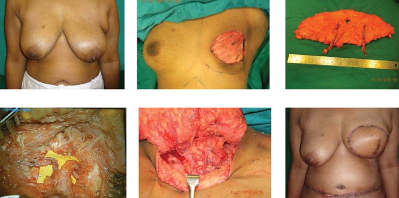
A bipedicled DIEP ([Fig. 7]) flap is harvested and needs two sets of microvascular anastomoses. The DIEP flap, in terms of volume and skin, needs to match the defect created by mastectomy or chest wall resection. In patients where the abdominal pannus is not so abundant or in cases when the requirement is large, the entire pannus and occasionally extra fat beyond the pannus needs to be harvested. To match the perfusion of this skin and volume of the flap, a bipedicle DIEP flap is often needed. In addition to this, need for a bipedicled flap could also be necessitated by a poor midline crossover perfusion, as per the native anatomy or due to midline/paramedian scars from previous surgery.
The method we used for the assessment of perfusion to judge the need for a bipedicle flap consisted of series of steps ([Fig. 8]). This was preceded by a CT angiogram in all cases planned for DIEP flap. The final decision was based on clinical judgement.
-
Large free ALT ([Fig. 9]) flaps or both free ALT and free tensor fascia latae (TFL) flaps as double free flaps are sometimes needed for radical mastectomy for recurrent or large primary invasive ductal carcinoma, malignant phyllodes, or rarer chest wall tumors.[2] Sometimes the large ALT flaps are perfused by multiple perforators arising from two separate pedicles. In case of combined free ALT and TFL flaps, their use as separate free flaps requires extra set of vessels for the other flap. To restore the perfusion of the flap in these cases adequately two sets of microvascular anastomoses are sometimes needed. The first donor pedicle of choice for these flaps is also IMA and V on the antegrade flow. The retrograde flow on the distal end can be used as the second donor vessel set.
-
When gracilis myocutaneous flap ([Fig. 10]) is used for breast reconstruction, sometimes there might be two vascular pedicles perfusing the muscle and overlying skin and subcutaneous tissue. In cases of doubtful perfusion from either of the pedicles, both pedicles can be anastomosed.
-
A single set of microvascular anastomoses might also need an alternative donor vessel in case of gross lymph nodal involvement, fibrosis, thrombosis, or injury precluding the use of the proximal IMA/V on antegrade flow.
-
A second venous microvascular anastomoses might be desired of the deep inferior epigastric vein pedicle or the superficial inferior epigastric vein. The distal end of IMV offers an easily available alternative for drainage.


The second set of microvascular anastomoses, when needed, can be done to a fresh set of donor vessels (supercharging) or to a branch or runoff of the first set of vessels (turbocharging). We prefer to supercharge as thrombosis of one vessel will not affect the other and the size discrepancy between a small branch and larger pedicle is taken care of.
The distal cut end of IMA/V with retrograde flow is an option, easily available, and accessible as field of dissection is same as that of the antegrade one. We chose retrograde limb as second donor set whenever feasible. The other choices for second pedicle were in the sequence: IM artery perforators, serratus branch of thoracodorsal vessel, thoracodorsal vessels, thoracoacromial vessels, lateral thoracic vessels, and transverse cervical vessels. Kanoi et al published their study on IMA perforators being consistent in anatomical location but their use in free tissue transfer for breast reconstruction depends on various other factors.[3] All these choices have some problem of either availability, accessibility, expendability, or caliber of vessel.
The use of the retrograde flow of IMA and V has evolved over the years in a fascinating way. Earliest use of retrograde limb arose with its anastomosis with posterior descending coronary artery during coronary artery bypass to relieve blockage in coronary vessels where antegrade vessel was anastomosed with left anterior descending vessels.[4] The anatomical basis of retrograde flow can be explained by the intricate arterial and venous communications of thoracic wall. The arterial network is derived mainly from thoracic aorta. Posterior intercostal and subcostal arteries from axillary artery, IMA from subclavian artery, and superior intercostal arteries anastomose permitting collateral circulation.[5] Anterior and posterior intercostal veins being separate vessels, normally draining in opposite directions, the tributaries of these veins anastomose approximately in anterior axillary line and due to lack of valves flow can be reversed.[5] [6]
In a case reported by Hassan et al, TRAM flap was used to perform breast reconstruction where intraoperatively due to fraility of antegrade IMA, the retrograde end of IMA was used for anastomosis and resulted in viable and well perfused flap thereby resulted as a salvage option.[7]
Li et al were the first to study the pressures in antegrade and retrograde limbs of IM arteries in dogs.[8] Adequacy of pressures flow in antegrade and retrograde limbs was established by Tomioka et al.[9] Further adequacy of flow in retrograde limbs was validated using intraoperative color Doppler and indocyanine green angiography by Kerr-Valentic et al[10] and Mohebali et al,[11] respectively. A case series of 10 flaps was performed to study the comparison of flow rates in antegrade and retrograde veins of IM vessels.[12]
Another case reported by Chan et al where in unilateral breast reconstruction with stacked DIEP flaps, both the antegrade and retrograde limbs were utilized for two flaps and they concluded that to increase the volume of reconstructed breast, retrograde limb of IMA can be used as second donor pedicle if stacking of flaps is done.[13] With this concept of retrograde set of IM vessels utilization, bilateral breast reconstruction was performed in two cases using single-sided recipient vessels where opposite-sided pedicle of DIEP was tuned in subcutaneous plane across the sternum and was anastomosed with retrograde set of IM vessels.[14] Another study done in 250 free flaps for bilateral breast reconstruction using single set of IM vessels by Opsomer et al documented the adequacy of retrograde limb without any significant predisposition of flap failure utilizing the same.[15] Retrograde limb of IM vessels is an invaluable option as second set of donor vessels and adequacy of venous flow can be justified by its valveless system.[6] Utilizing the retrograde flow does not consume any extra time in exposing and dissecting the vessels as it is already done for the proximal limb antegrade flow.
Most of the breast reconstructions where a bipedicled flap was done and no thrombotic event happened were uneventful in the late postoperative period with no fat necrosis. These cases if harvested on a single pedicle would be very high risk for partial necrosis or fat necrosis.
No imaging was performed to document the patency of vessels. The partial flap necrosis complications could be attributed to either thrombosis of one of the microvascular anastomoses or insufficient drainage of a patent microvascular anastomoses.
We do not have a comparator arm as all cases where perfusion was suspect additional pedicles were anastomosed.
Conclusion
The retrograde flow of the distal cut end of IMA and IMV can be safely used as a donor vessel for microvascular anastomoses for free tissue transfer for breast reconstruction or chest wall coverage. The benefit is that it can be safely used for supercharging, superdrainage, or as the sole donor for microvascular anastomoses in event when proximal flow IMA/V is unavailable due to gross lymph nodal involvement, fibrosis, thrombosis, or injury without any consumption of time in dissection of other set of vessels in the vicinity. Though it might appear counterintuitive, the blood flow rates and direction are not an impediment to the use of distal cut ends of IMA or IMV. The only disadvantage that appears is the unavailability of IMA/V for coronary artery bypass in cases where patient develops coronary heart disease in later age.
Conflict of Interest
None.
-
References
- 1 Sung H, Ferlay J, Siegel RL. et al. Global Cancer Statistics 2020: GLOBOCAN Estimates of Incidence and Mortality Worldwide for 36 Cancers in 185 Countries. CA Cancer J Clin 2021; 71 (03) 209-249
- 2 Jaiswal D, Mantri MR, Shankhdhar VK, Wagh SH. Chimeric ALT Plus TFL perforator flap for breast reconstruction post radical mastectomy with large skin defect. Indian J Plast Surg 2021; 54 (02) 208-210
- 3 Kanoi AV, Panchal KB, Sen S, Biswas G. Computed tomography angiographic study of internal mammary perforators and their use as recipient vessels for free tissue transfer in breast reconstruction. Indian J Plast Surg 2017; 50 (01) 50-55
- 4 Goiti JJ, Smith GH. Coronary artery surgery using inverted internal mammary artery. Br Heart J 1982; 48 (01) 81-82
- 5 Clinically Oriented Anatomy. 5th edition. Philadelphia: Lippincott Williams & Wilkins; 2006. :103–104
- 6 Al-Dhamin A, Bissell MB, Prasad V, Morris SF. The use of retrograde limb of internal mammary vein in autologous breast reconstruction with DIEAP flap: anatomical and clinical study. Ann Plast Surg 2014; 72 (03) 281-284
- 7 Hassan S, Rasheed T, Raurell A. Salvage of a TRAM breast reconstruction flap using the retrograde internal mammary artery system. Indian J Plast Surg 2014; 47 (03) 447-449
- 8 Li S, Mu L, Li Y. et al. Breast reconstruction with the free bipedicled inferior TRAM flap by anastomosis to the proximal and distal ends of the internal mammary vessels. J Reconstr Microsurg 2002; 18 (03) 161-168
- 9 Tomioka YK, Uda H, Yoshimura K, Sunaga A, Kamochi H, Sugawara Y. Studying the blood pressures of antegrade and retrograde internal mammary vessels: do they really work as recipient vessels?. J Plast Reconstr Aesthet Surg 2017; 70 (10) 1391-1396
- 10 Kerr-Valentic MA, Gottlieb LJ, Agarwal JP. The retrograde limb of the internal mammary vein: an additional outflow option in DIEP flap breast reconstruction. Plast Reconstr Surg 2009; 124 (03) 717-721
- 11 Mohebali J, Gottlieb LJ, Agarwal JP. Further validation for use of the retrograde limb of the internal mammary vein in deep inferior epigastric perforator flap breast reconstruction using laser-assisted indocyanine green angiography. J Reconstr Microsurg 2010; 26 (02) 131-135
- 12 Venturi ML, Poh MM, Chevray PM, Hanasono MM. Comparison of flow rates in the antegrade and retrograde internal mammary vein for free flap breast reconstruction. Microsurgery 2011; 31 (08) 596-602
- 13 Chan RK, Przylecki W, Guo L, Caterson SA. Case report. The use of both antegrade and retrograde internal mammary vessels in a folded, stacked deep inferior epigastric artery perforator flap. Eplasty 2010; 10: e32
- 14 Lee JH, Varon DE, Halvorson EG. Unilateral internal mammary recipient vessels for bilateral DIEP flap breast reconstruction. Plast Reconstr Surg Glob Open 2017; 5 (06) e1359
- 15 Opsomer D, D’Arpa S, Benmeridja L, Stillaert F, Noel W, Van Landuyt K. Bilateral DIEP flap breast reconstruction to a single set of Internal Mammary vessels: technique, safety, and outcomes after 250 flaps. Plast Reconstr Surg 2019; 144 (04) 554e-564e
Address for correspondence
Publication History
Article published online:
14 July 2022
© 2022. Association of Plastic Surgeons of India. This is an open access article published by Thieme under the terms of the Creative Commons Attribution-NonDerivative-NonCommercial License, permitting copying and reproduction so long as the original work is given appropriate credit. Contents may not be used for commercial purposes, or adapted, remixed, transformed or built upon. (https://creativecommons.org/licenses/by-nc-nd/4.0/)
Thieme Medical and Scientific Publishers Pvt. Ltd.
A-12, 2nd Floor, Sector 2, Noida-201301 UP, India
-
References
- 1 Sung H, Ferlay J, Siegel RL. et al. Global Cancer Statistics 2020: GLOBOCAN Estimates of Incidence and Mortality Worldwide for 36 Cancers in 185 Countries. CA Cancer J Clin 2021; 71 (03) 209-249
- 2 Jaiswal D, Mantri MR, Shankhdhar VK, Wagh SH. Chimeric ALT Plus TFL perforator flap for breast reconstruction post radical mastectomy with large skin defect. Indian J Plast Surg 2021; 54 (02) 208-210
- 3 Kanoi AV, Panchal KB, Sen S, Biswas G. Computed tomography angiographic study of internal mammary perforators and their use as recipient vessels for free tissue transfer in breast reconstruction. Indian J Plast Surg 2017; 50 (01) 50-55
- 4 Goiti JJ, Smith GH. Coronary artery surgery using inverted internal mammary artery. Br Heart J 1982; 48 (01) 81-82
- 5 Clinically Oriented Anatomy. 5th edition. Philadelphia: Lippincott Williams & Wilkins; 2006. :103–104
- 6 Al-Dhamin A, Bissell MB, Prasad V, Morris SF. The use of retrograde limb of internal mammary vein in autologous breast reconstruction with DIEAP flap: anatomical and clinical study. Ann Plast Surg 2014; 72 (03) 281-284
- 7 Hassan S, Rasheed T, Raurell A. Salvage of a TRAM breast reconstruction flap using the retrograde internal mammary artery system. Indian J Plast Surg 2014; 47 (03) 447-449
- 8 Li S, Mu L, Li Y. et al. Breast reconstruction with the free bipedicled inferior TRAM flap by anastomosis to the proximal and distal ends of the internal mammary vessels. J Reconstr Microsurg 2002; 18 (03) 161-168
- 9 Tomioka YK, Uda H, Yoshimura K, Sunaga A, Kamochi H, Sugawara Y. Studying the blood pressures of antegrade and retrograde internal mammary vessels: do they really work as recipient vessels?. J Plast Reconstr Aesthet Surg 2017; 70 (10) 1391-1396
- 10 Kerr-Valentic MA, Gottlieb LJ, Agarwal JP. The retrograde limb of the internal mammary vein: an additional outflow option in DIEP flap breast reconstruction. Plast Reconstr Surg 2009; 124 (03) 717-721
- 11 Mohebali J, Gottlieb LJ, Agarwal JP. Further validation for use of the retrograde limb of the internal mammary vein in deep inferior epigastric perforator flap breast reconstruction using laser-assisted indocyanine green angiography. J Reconstr Microsurg 2010; 26 (02) 131-135
- 12 Venturi ML, Poh MM, Chevray PM, Hanasono MM. Comparison of flow rates in the antegrade and retrograde internal mammary vein for free flap breast reconstruction. Microsurgery 2011; 31 (08) 596-602
- 13 Chan RK, Przylecki W, Guo L, Caterson SA. Case report. The use of both antegrade and retrograde internal mammary vessels in a folded, stacked deep inferior epigastric artery perforator flap. Eplasty 2010; 10: e32
- 14 Lee JH, Varon DE, Halvorson EG. Unilateral internal mammary recipient vessels for bilateral DIEP flap breast reconstruction. Plast Reconstr Surg Glob Open 2017; 5 (06) e1359
- 15 Opsomer D, D’Arpa S, Benmeridja L, Stillaert F, Noel W, Van Landuyt K. Bilateral DIEP flap breast reconstruction to a single set of Internal Mammary vessels: technique, safety, and outcomes after 250 flaps. Plast Reconstr Surg 2019; 144 (04) 554e-564e