
Subscribe to RSS
DOI: 10.1055/s-0041-1731419
Anatomical Study of the Medial Region of the Knee: Qualitative, Quantitative Analysis and Description of the Anterior Oblique Ligament
Article in several languages: português | English
Abstract
Objective To describe all ligamentous, capsular, tendinous and bone landmarks structures of the medial region of the knee, as well as a new ligamentous structure identified in a series of anatomical dissections of cadaveric specimens.
Methods Twenty cadaver knees were dissected to study the medial compartment. The main structures of this region were identified during dissection. The morphology of the structures and their relationship with known anatomical parameters were determined both qualitatively and quantitatively. The collected data were analyzed and interpreted using descriptive statistics.
Results In the dissection of all specimens, all ligamentous structures previously described in the anatomical study of the medial part of the knee were identified, and objective measures that can help as parameters for surgical ligament reconstruction were identified. When dissecting the medial collateral ligament, a bony prominence immediately distal to its proximal tibial insertion was observed and described, as well as a bursa below the ligament, in which it was not inserted. We also described a ligamentous structure with extracapsular location, originated anteriorly to the medial epicondyle and following obliquely towards the tibia. These structures were named, respectively, interinsertional tubercle, interinsertional bursa and anterior oblique ligament.
Conclusion In addition to the description and measurement of the structures and parameters already existing in the anatomical study of the medial part of the knee, it was possible to describe three new structures not yet described in the literature: the interinsertional tubercle, the interinsertional bursa, and the anterior oblique ligament. These structures were found in all dissections performed.
Keywords
medial collateral ligament, knee/anatomy & histology - anterior cruciate ligament reconstruction - anterior cruciate ligament injuries - anterior cruciate ligament - joint instabilityIntroduction
The medial structures of the knee play a primary role in stabilizing valgus stress, as well as rotational stress, which makes them extremely important in the overall evaluation of the knee with ligament injury and in the choice of the most appropriate treatment. The medial collateral ligament (MCL) is the most commonly injured knee ligament.[1] However, the importance given to the anatomical study of these structures was small compared with the lateral portion, which can be explained by the fact that the MCL has a great potential for healing after injury.[2]
Warren et al.[3] described the medial anatomy of the knee with a division into three distinct layers, a concept still used nowadays Layer 1 is composed of the crural fascia, from the patella to the popliteal fossa, including the fascia of the sartorius. Between layers 1 and 2, are the gracilis and semitendinosus tendons. Layer 2, is composed of the superficial medial collateral ligament (MCLs). The third layer consists of the joint capsule and the deep MCL.
Years after that description, Robinson et al.[4] published a new anatomical study with a new division of the medial portion of the knee into three parts, from anterior to posterior. The most anterior part comprises the structures of the medial limit of the patellar tendon to the anterior edge of the longitudinal fibers of the MCLs. The middle part encompasses the MCLs itself, and the posterior part extends from the posterior edge of the longitudinal fibers of the MCLs to the medial head of the gastrocnemius.[2] LaPrade et al.,[5] in an article published in 2007, brought the concept that simple division by layers, although adequate, may not be the best method of anatomical description, since knowledge of the origins and insertions of structures is essential for the planning of a surgical reconstruction. The present study focused on the quantitative evaluation of the medial part of the knee, different from previous studies that focused on simple qualitative description. Thus, it presented consistent patterns of measures that could be applied in the planning of surgical reconstructions.
With this relatively recent history of studies,[4] [6] it is possible to note the importance of the anatomical study of the medial part of the knee in the search for a surgical ligament reconstruction most similar to the original anatomical condition. In the present study, we attempted to describe and guide the anatomical dissection of the medial part of the knee, step by step, to identify all structures with biomechanical importance, as well as their origins and insertions, with precise, practical, and objective measurements. In addition, it was possible to describe new anatomical structures, not yet found in the literature, which may be of great importance in the biomechanical study of the knee.
Materials and methods
Between April and August 2020, 20 knees derived from transfemoral amputations performed exclusively for vascular reasons and without signs of trauma were dissected. The following exclusion criteria were applied: signs of trauma, previous surgery, macroscopic signs of osteoarthritis, and poor state of preservation. Thus, 19 knees were studied. The knees were stored in 10% formaldehyde solution and refrigerated at 5.3°C. Dissections were performed 2 to 21 days after amputation. The present study was approved by the research ethics committee of the institution.
The specimens were photographed in a 12-megapixel digital camera and were measured in a digital caliper with an accuracy of 0.01 mm (Mitutoyo Sul Americana Ltda. - Suzano, SP - Brasil).
All specimens were identified according to gender, age, dissected side, amputation date, and date of dissection. The collected data were analyzed and interpreted using descriptive statistics.
Results
A curvilinear hockey-stick incision of ∼ 25 cm was made following a line that cuts the femoral diaphysis, the medial epicondyle and the long axis of the tibia, starting 4 cm proximal to the medial epicondyle and extending to the base of the insertion of the pes anserinus, with a gentle opening of the dermis and of the subcutaneous cellular tissue until the exposure of the nacreous coloring that corresponds to a bright fascia, which we called medial bone fascia. Extensive dissection was performed in the subcutaneous plane until complete exposure of the fascia from anteromedial to posteromedial ([Figure 1]).

At the level of the popliteal fossa, the fascia recess, widely covered by adipose tissue, was observed, and careful resection of this tissue was performed until the exposure of the posteromedial tendinous structures ([Figure 2]).

The most delicate point of dissection is the plane below the fascia, which is closely adhered to all deeper structures, demanding substantial care not to damage the structures of interest. Through a superficial longitudinal incision, the fascia is divulsioned, creating a space between it and the other structures ([Figure 3]).
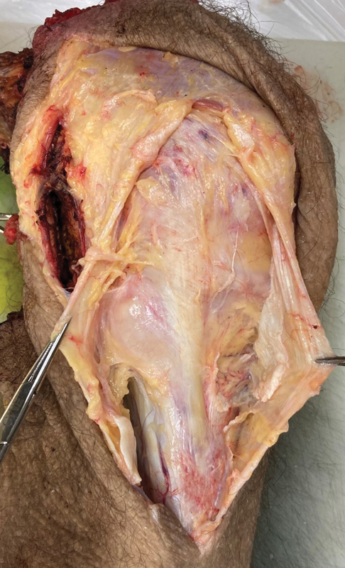
The adductor magnus tendon inserted to a posterior bone depression proximal to the tubercle of the adductor was identified. The medial distal aspect of the adductor magnus tendon presented a thick fascial expansion, which spread posteromedially. Its oval-shaped insertion averaged 9.38 × 9.82 mm [(Figure 4).]

The insertion of the medial gastrocnemius tendon was proximal and posterior in a depression adjacent to the gastrocnemius tubercle and distal and posterior to the adductor tubercle. As previously observed, the medial gastrocnemius tendon presented a thick fascial bond along its lateral face, distancing on average 10.92 mm from the adductor magnus tendon.
The medial patellofemoral ligament was located anteriorly and in a distinct extra-articular layer of the medial joint capsule, and it was identified from the distal edge of the vastus medialis oblique. Its insertion in the femur was proximal and posterior to the medial and anterior epicondyle and distal to the adductor tubercle. The mean length of the medial patellofemoral ligament was 50.52 mm ([Figure 5]).

The MCLs is inserted in the femur in a small depression, slightly proximal and posterior to the center of the medial epicondyle, ∼ 30.71 mm from the articular surface, anterior and distal to the medial gastrocnemius and adductor magnus tendons. The MCLs presented two distinct tibial insertions; the distal tibial insertion was closely adhered to the bone and the periosteum, being possible to fully visualize it after identification and resection of the tendons of the pes anserinus, and it was located at an average of 66.85 mm of the joint line ([Figure 6]).

Distal disinsertion of the periosteum was performed until a bursa was visualized below the ligament, where it was not inserted into the bone. We named this structure the interinsertional bursa. Proximally to the bursa, there was a bony protrusion located at an average of 21.35 mm from the articular surface and 28.35 mm from the distal insertion of the superficial medial collateral ligament. We named this structure an interinsertional tubercle. Proximal to it, sets in the proximal tibial insertion of the MCLs, at an average distance of 12.45 mm from the joint line ([Figure 7]).

With the disinsertion of the MCL, it is possible to visualize a characteristic ligamentous structure, whose origin is anterior to the origin of the superficial medial collateral ligament and runs with oblique fibers towards the tibia with insertion at ∼ 9.11 mm anterior to the anterior edge of the MCLs, in a fan shape ([Figure 8]).

This structure, which was named oblique anterior ligament, was more evident after release of the MCLs and through external rotation of the tibia. It had an average length of 33.82 mm in flexion of 90°and of 26.56 mm in full extension, with an average width of 6.83 mm at the origin, 13.06 mm at insertion, and 8.05 mm at the level of the joint line.
The deep MCL has its femoral insertion with an oval shape in the anteroposterior extension, and proximal to the articular cartilage of the femur, immediately posterior and distal to the fixation of the MCLs and on the lower surface of the medial epicondyle.
The tibial insertion was distal to the margin of the articular cartilage, immediately proximal to the insertion of the semimembranosus tendon. The fibers were parallel and flowed distally and slightly anteriorly. Although it was a distinct structure, it was inseparable from the capsule and was also fixed to the medial meniscus. The anterior edge was easily identifiable, as the deep MCL formed an easily palpable thick band within the surrounding capsular tissue. Posteriorly, the proximal portion of the ligament was mixed with the posteromedial capsule.
Discussion
The anatomy of the medial region of the knee is very important because its structures are responsible for the primary stabilization against the valgus and external rotation forces applied to the knee.[2] [3] The lesion of the MCL is the most common among the ligament injuries associated with anterior cruciate ligament injury, which makes the present study even more relevant.
However, in the literature, the importance of these structures is neglected when compared with that of lateral structures and of central pivot structures. A probable reason for this is the high healing potential of the MCL, despite the high injury rates.[2] [7] [8] Another possible cause comes from the difficulty of dissection of the medial components of the knee, since there is great adherence to the fascial planes and a thick overlap of connective tissues, which makes the solated delimitation of structures difficult and laborious. For this reason, the descriptions of qualitative fixation parameters of medial ligament reconstruction of the knee are imprecise.
In most anatomy reviews, three bony prominences were always found (gastrocnemius tubercle, adductor tubercle, and medial epicondyle)[9] [10] ([Figure 9]). However, no description of the tibial bony prominence that lies below the MCLs immediately distal to its proximal tibial insertion – the interinsertional tubercle – as well as the bursa that is in the same topography – the interinsertional bursa – was found. The probable cause for this is the difficulty in disinsertion of the MCLs and its adherence to the medial bone fascia, which hinders its dissection and isolation. Such parameters may become of paramount importance in the advancement of medial ligament reconstruction techniques of the knee, since they are structures immediately distal to the proximal tibial insertion of the MCLs.

The MCLs, in its femoral origin, was described as anterior and distal to the adductor tubercle in an oval shape.[11] [12] The distal tibial insertion is between 46 and 60 mm distal to the joint,[13 ]of which its total length is 90 to 110 mm.[5] [14] In its distal tibial insertion, the fibers mix tangentially with the periosteum.[15] [16] The MCLs presented, in the present study, with an average width of 11.35 mm in its femoral insertion, presenting in its distal tibial insertion an average width of 12.30 mm, with fibers connected to the pes anserinus.[17] It is described as having parallel fibers and oblique fibers, with a fan-shaped expansion of connective tissues extending from its anterior edge towards the patella, mixing with the medial patellar retinaculum,[18] [19] which is observed in ∼ 42% of the dissections.
Most authors agree that the deep MCL is adhered to the medial meniscus and is confused with the meniscofemoral and meniscotibial ligaments,[7 ]being described as inserted in the tibia immediately distal to its articular surface or, in some cases, in the articular margin of the tibia.[20] [21] There is disagreement, however, regarding the differentiation of the deep MCL from the joint capsule. In some cases, it is described as continuous to the capsule, mixing with medial capsular fibers. In other knees, the deep MCL was identified as a separate structure of the capsule and was, therefore, not adhered to the meniscus.[22] There is also a description of the femoral meniscus ligament as part of the deep MCL and in other sections, as a distinct structure.[15] [23] In our dissection, all specimens presented the ligament and the capsule as inseparable structures, and their isolation was not possible.
During the dissections, a ligament structure of oblique orientation not yet described in the literature, between the femur and tibia, was observed. It was found in 100% of the pieces. Its location was extracapsular and easily distinguishable from the joint capsule ([Figure 9]). It was named the anterior oblique ligament, and it is described in detail in a study entirely dedicated to it, in which its existence is evidenced in radiological studies with magnetic resonance imaging (MRI) and histological studies that confirm the presence of ligament tissue. Its femoral origin occurred directly on the anterior face of the medial epicondyle, confusing with the femoral insertion of the MCLs. Some reasons were raised to justify the fact that this structure has not been described previously. One of them is the use of the Warren layered description for a long period, which has proven to be ineffective in medial dissection of the knee and in the visualization of structures in isolation. Another justification would be the difficulty to visualize the structure, which is only evident after the release of the whole MCLs ([Figure 10]).

The finding of this new ligament structure allows a new view in relation to circumferential joint structures and rotational control, which we named Quadrant Theory. This means, in a summarized way, that the structures found in the anteromedial quadrant of the knee (delimited by the transepicondilar axis and a line perpendicular to the transverse articular axis) would be responsible for the valgus restriction and external rotation. This new perspective on rotational control can help to evaluate the need for peripheral reconstructions complementary to the central pivot that would promote greater stability and lower failure rate.
Conclusion
In the present study, a qualitative and quantitative description of the main medial structures of the knee was performed, as well as that of three new structures not yet described in the literature: the interinsertional tubercle and bursa, and the oblique anterior ligament. With these findings and with a better evaluation of the entire medial anatomy of the knee, which has sometimes been neglected in previous anatomical studies, it is believed that it will be possible to improve existing ligament reconstruction techniques, as well as to more fully understand knee biomechanics. Thus, it would become possible to plan a surgical reconstruction most similar to the original anatomical condition, improving the postoperative outcome and decreasing the failure rate.
Conflito de Interesses
Os autores declaram não haver conflito de interesses.
Note
Work developed at the Irmandade da Santa Casa de Misericórdia de São Paulo, Faculdade de Ciências Médicas, São Paulo, SP, Brazil
-
Referências
- 1 LaPrade RF. The medial collateral ligament complex and the posterolateral aspect of the knee. In: Arendt EA. editor. Orthopaedic knowledge update. Sports medicine 2.. Rosemont, IL: American Academy of Orthopaedic Surgeons; 1999: 327-340
- 2 Warren LA, Marshall JL, Girgis F. The prime static stabilizer of the medical side of the knee. J Bone Joint Surg Am 1974; 56 (04) 665-674
- 3 Warren LF, Marshall JL. The supporting structures and layers on the medial side of the knee: an anatomical analysis. J Bone Joint Surg Am 1979; 61 (01) 56-62
- 4 Robinson JR, Bull AM, Thomas RR, Amis AA. The role of the medial collateral ligament and posteromedial capsule in controlling knee laxity. Am J Sports Med 2006; 34 (11) 1815-1823
- 5
LaPrade RF,
Morgan PM,
Wentorf FA,
Johansen S,
Engebretsen L.
The anatomy of the posterior aspect of the knee. An anatomic study. J Bone Joint Surg
Am 2007; 89 (04) 758-764
MissingFormLabel
- 6
LaPrade RF,
Engebretsen AH,
Ly TV,
Johansen S,
Wentorf FA,
Engebretsen L.
The anatomy of the medial part of the knee. J Bone Joint Surg Am 2007; 89 (09) 2000-2010
MissingFormLabel
- 7 Loredo R, Hodler J, Pedowitz R, Yeh LR, Trudell D, Resnick D. Posteromedial corner of the knee: MR imaging with gross anatomic correlation. Skeletal Radiol 1999; 28 (06) 305-311
- 8 Fanelli GC, Harris JD. Surgical treatment of acute medial collateral ligament and posteromedial corner injuries of the knee. Sports Med Arthrosc Rev 2006; 14 (02) 78-83
- 9 Kaplan EB. Factors responsible for the stability of the knee joint. Bull Hosp Jt Dis 1957; 18 (01) 51-59
- 10 Poliacu Prosé L, Lohman AH, Huson A. The collateral ligaments of the knee joint in the cat and man. Morphological and functional study of the internal arrangement of fibers. Acta Anat (Basel) 1988; 133 (01) 70-78
- 11 Slocum DB, Larson RL. Rotatory instability of the knee. Its pathogenesis and a clinical test to demonstrate its presence. J Bone Joint Surg Am 1968; 50 (02) 211-225
- 12 Tuxøe JI, Teir M, Winge S, Nielsen PL. The medial patellofemoral ligament: a dissection study. Knee Surg Sports Traumatol Arthrosc 2002; 10 (03) 138-140
- 13 Ivey M, Prud'homme J. Anatomic variations of the pes anserinus: a cadaver study. Orthopedics 1993; 16 (05) 601-606
- 14 Cohen M, Astur DC, Branco RC. et al. An anatomical three-dimensional study of the posteromedial corner of the knee. Knee Surg Sports Traumatol Arthrosc 2011; 19 (10) 1614-1619
- 15 Brantigan OC, Voshell AF. The tibial collateral ligament: its function, its bursae, and its relation to the medial meniscus. J Bone Joint Surg Am 1943; 25 (01) 121-131
- 16
Hughston JC,
Andrews JR,
Cross MJ,
Moschi A.
Classification of knee ligament instabilities. Part I. The medial compartment and
cruciate ligaments. J Bone Joint Surg Am 1976; 58 (02) 159-172
MissingFormLabel
- 17 Sims WF, Jacobson KE. The posteromedial corner of the knee: medial-sided injury patterns revisited. Am J Sports Med 2004; 32 (02) 337-345
- 18 Steensen RN, Dopirak RM, McDonald III WG. The anatomy and isometry of the medial patellofemoral ligament: implications for reconstruction. Am J Sports Med 2004; 32 (06) 1509-1513
- 19 Amis AA, Firer P, Mountney J, Senavongse W, Thomas NP. Anatomy and biomechanics of the medial patellofemoral ligament. Knee 2003; 10 (03) 215-220
- 20 Standring S. Editor. Gray's anatomy: the anatomical basis of clinical practice. 39th ed.. New York: Churchill Livingstone; 2005
- 21
De Maeseneer M,
Van Roy F,
Lenchik L,
Barbaix E,
De Ridder F,
Osteaux M.
Three layers of the medial capsular and supporting structures of the knee: MR imaging-anatomic
correlation. Radiographics 2000; 20 (Spec No): S83-S89
MissingFormLabel
- 22 Thompson JC. Editor. Netter's concise atlas of orthopaedic anatomy. Teterboro, NJ: Icon Learning Systems; 2002
- 23 Brantigan OC, Voshell AF. The mechanics of the ligaments and menisci of the knee joint. J Bone Joint Surg Am 1941; 23: 44-66
Endereço para correspondência
Publication History
Received: 01 October 2020
Accepted: 08 March 2021
Article published online:
13 October 2021
© 2021. Sociedade Brasileira de Ortopedia e Traumatologia. This is an open access article published by Thieme under the terms of the Creative Commons Attribution-NonDerivative-NonCommercial License, permitting copying and reproduction so long as the original work is given appropriate credit. Contents may not be used for commecial purposes, or adapted, remixed, transformed or built upon. (https://creativecommons.org/licenses/by-nc-nd/4.0/)
Thieme Revinter Publicações Ltda.
Rua do Matoso 170, Rio de Janeiro, RJ, CEP 20270-135, Brazil
-
Referências
- 1 LaPrade RF. The medial collateral ligament complex and the posterolateral aspect of the knee. In: Arendt EA. editor. Orthopaedic knowledge update. Sports medicine 2.. Rosemont, IL: American Academy of Orthopaedic Surgeons; 1999: 327-340
- 2 Warren LA, Marshall JL, Girgis F. The prime static stabilizer of the medical side of the knee. J Bone Joint Surg Am 1974; 56 (04) 665-674
- 3 Warren LF, Marshall JL. The supporting structures and layers on the medial side of the knee: an anatomical analysis. J Bone Joint Surg Am 1979; 61 (01) 56-62
- 4 Robinson JR, Bull AM, Thomas RR, Amis AA. The role of the medial collateral ligament and posteromedial capsule in controlling knee laxity. Am J Sports Med 2006; 34 (11) 1815-1823
- 5
LaPrade RF,
Morgan PM,
Wentorf FA,
Johansen S,
Engebretsen L.
The anatomy of the posterior aspect of the knee. An anatomic study. J Bone Joint Surg
Am 2007; 89 (04) 758-764
MissingFormLabel
- 6
LaPrade RF,
Engebretsen AH,
Ly TV,
Johansen S,
Wentorf FA,
Engebretsen L.
The anatomy of the medial part of the knee. J Bone Joint Surg Am 2007; 89 (09) 2000-2010
MissingFormLabel
- 7 Loredo R, Hodler J, Pedowitz R, Yeh LR, Trudell D, Resnick D. Posteromedial corner of the knee: MR imaging with gross anatomic correlation. Skeletal Radiol 1999; 28 (06) 305-311
- 8 Fanelli GC, Harris JD. Surgical treatment of acute medial collateral ligament and posteromedial corner injuries of the knee. Sports Med Arthrosc Rev 2006; 14 (02) 78-83
- 9 Kaplan EB. Factors responsible for the stability of the knee joint. Bull Hosp Jt Dis 1957; 18 (01) 51-59
- 10 Poliacu Prosé L, Lohman AH, Huson A. The collateral ligaments of the knee joint in the cat and man. Morphological and functional study of the internal arrangement of fibers. Acta Anat (Basel) 1988; 133 (01) 70-78
- 11 Slocum DB, Larson RL. Rotatory instability of the knee. Its pathogenesis and a clinical test to demonstrate its presence. J Bone Joint Surg Am 1968; 50 (02) 211-225
- 12 Tuxøe JI, Teir M, Winge S, Nielsen PL. The medial patellofemoral ligament: a dissection study. Knee Surg Sports Traumatol Arthrosc 2002; 10 (03) 138-140
- 13 Ivey M, Prud'homme J. Anatomic variations of the pes anserinus: a cadaver study. Orthopedics 1993; 16 (05) 601-606
- 14 Cohen M, Astur DC, Branco RC. et al. An anatomical three-dimensional study of the posteromedial corner of the knee. Knee Surg Sports Traumatol Arthrosc 2011; 19 (10) 1614-1619
- 15 Brantigan OC, Voshell AF. The tibial collateral ligament: its function, its bursae, and its relation to the medial meniscus. J Bone Joint Surg Am 1943; 25 (01) 121-131
- 16
Hughston JC,
Andrews JR,
Cross MJ,
Moschi A.
Classification of knee ligament instabilities. Part I. The medial compartment and
cruciate ligaments. J Bone Joint Surg Am 1976; 58 (02) 159-172
MissingFormLabel
- 17 Sims WF, Jacobson KE. The posteromedial corner of the knee: medial-sided injury patterns revisited. Am J Sports Med 2004; 32 (02) 337-345
- 18 Steensen RN, Dopirak RM, McDonald III WG. The anatomy and isometry of the medial patellofemoral ligament: implications for reconstruction. Am J Sports Med 2004; 32 (06) 1509-1513
- 19 Amis AA, Firer P, Mountney J, Senavongse W, Thomas NP. Anatomy and biomechanics of the medial patellofemoral ligament. Knee 2003; 10 (03) 215-220
- 20 Standring S. Editor. Gray's anatomy: the anatomical basis of clinical practice. 39th ed.. New York: Churchill Livingstone; 2005
- 21
De Maeseneer M,
Van Roy F,
Lenchik L,
Barbaix E,
De Ridder F,
Osteaux M.
Three layers of the medial capsular and supporting structures of the knee: MR imaging-anatomic
correlation. Radiographics 2000; 20 (Spec No): S83-S89
MissingFormLabel
- 22 Thompson JC. Editor. Netter's concise atlas of orthopaedic anatomy. Teterboro, NJ: Icon Learning Systems; 2002
- 23 Brantigan OC, Voshell AF. The mechanics of the ligaments and menisci of the knee joint. J Bone Joint Surg Am 1941; 23: 44-66


