Introduction
Smoking is clearly associated with the severity of periodontal disease, since evidence suggests that smokers showed high incidence of chronic periodontal disease and loss of clinical attachment. Smokers were four times more likely to have periodontitis, compared with nonsmokers. Current smokers have deeper probing depths, greater attachment loss, greater bone loss, and fewer teeth.[1] Moreover, meta-analysis suggests that smoking has some effect on periodontal therapy, for both nonsurgical and surgical therapies. Periodontal pocket healing was slower for smoker than for nonsmoker patients.[2] In addition, smoking affects periodontal regeneration therapy, and in furcation grade II, significantly decreased defect fill was observed in smokers, when using bone graft and membrane.[3] Nicotine can also be detected in saliva, serum, and gingival crevice in smokers, where serum levels of nicotine after smoking the first cigarette and the second cigarette were 19 ± 11.3 ng/mL (0.117 ± 0.070 µM) and 22.9 ± 11.2 ng/mL (0.141 ± 0.069 µM), respectively.[4] A previous study has reported that concentrations of nicotine at 5 to 25 mM show toxicity to the periodontal ligament (PDL) cell in Dulbecco’s modified Eagle’s medium (DMEM) media.[5]
Porphyromonas gingivalis has been suggested to be a key periodontopathic bacteria, found in the subgingival plaque of smokers.[6]
[7]
Porphyromonas gingivalis was also shown to stimulate human dental follicle stem cells to produce cytokine, such as interleukin-8, which is related to inflammation.[8] The components of P. gingivalis involved in pathogenicity consists of lipopolysaccharide (LPS), fimbria, and bacterial toxin. LPS from P. gingivalis could inhibit proliferation and differentiation of mesenchymal stem cells (MSCs).[9] Moreover, LPS was shown to suppress the osteoblastic activity of PDL cells by promoting secretion of proinflammatory cytokine.[10] In addition, subgingival microbial flora were found to have higher levels of P. gingivalis in smokers than in nonsmokers, but without significant difference.[6]
[7]
PDL fibroblasts are very important in tissue regeneration, since they can undergo differentiation into several types of cells including osteoblasts. Osteogenic differentiation is associated with the expression of several genes, such as collagen type I (COL1A1), alkaline phosphatase (ALP), Runt-related transcription factor 2 (Runx2), and osteocalcin (OCN). COL1A1 is an important component of bone extracellular matrix, forming connections with cell surface integrins and other extracellular matrix proteins.[11] It also plays a role in cell adhesion, proliferation, and differentiation of the osteoblast phenotype. Runx2 is a major regulator of osteoblast differentiation. ALP was reported to act as an early indicator of osteoblast activity during early osteoblast. Levels of ALP mRNA have been shown to increase within 2 days after stimulation, followed by steady increases with the progression of osteoblastic differentiation, up to 14 days.[12] The OCN gene is one of the few osteoblast-specific genes, and encodes OCN, one of the most abundant proteins present in bone.[13] It plays an important role in the differentiation of osteoblast progenitor cells, and shows significant upregulation in both matrix synthesis and mineralization. Differentiation of PDL cells was found to be associated with the expression of COL1A1, ALP, Runx2, and OCN.[13] Nicotine also significantly decreased gene expression of ALP, OCN, and Runx2 during the osteogenic induction of the PDL stem cell.[14]
Currently, there is no information on the interaction between P. gingivalis and nicotine on the differentiation potential of PDL stem cells. Therefore, the present in vitro study was aimed at examining the osteogenic properties of PDL cells in the presence and absence of periodontal pathogens, with and without nicotine, under physiological conditions. We hope that this knowledge will be useful for the treatment of periodontitis in smoking patients, as well as to explain why periodontitis patients should refrain from smoking.
Materials and Methods
PDL Cell Culture
Human PDL cells from Science Cell Research Laboratories (Cat #2630; Science Cell Research Laboratories, Carlsbad, California, United States) were used in this study. Human PDL cells were cultured in DMEM (Gibco BRL, Long Island, New York, United States) and supplemented with 10% fetal bovine serum (FBS, Hyclone, Hyclone UK Ltd., UK), and 1% antibiotics (amphotericin B [25 µg/mL], streptomycin sulphate [10,000 µg/mL], and penicillin G [10,000 U/mL]), (Hyclone, Hyclone Laboratories, Inc., Utah, United States), then incubated in 5% CO2, 37°C, and 100% humidity. Cells at passage 4 to 6 was used for experiments.
Preparation of Porphyromonas gingivalis Lysate
Porphyromonas gingivalis (strain W50) obtained from American type culture collection (ATCC number 53978TM) was used in this study. Porphyromonas gingivalis was cultured in Trypticase soy broth in an anaerobic jar. Cells were harvested by centrifugation for 15 minutes at 2,060× g at 4°C, washed twice with PBS, and confirmed for purity by Gram staining. Cells were sonicated with high-intensity ultrasonication for 30 minutes. After that they were examined under a microscope to ensure complete cell breakdown, then centrifuged at 8,000× g for 10 minutes at 4°C to pellet cell debris. The sample cell lysate was filtered with a filter-sterilized unit (0.2 µm) and kept frozen at 80°C until used. Porphyromonas gingivalis lysate solution at 10, 20, 40, and 50 µg/mL were tested on PDLs.
Nicotine Preparation
Nicotine was purchased from Sigma-Aldrich, St Louis, Missouri, United States (nicotine 99% [TLC], liquid, N0267). Nicotine solutions was prepared into 200, 100, 50, 25, 20, 10, 1, 0.1, and 1 µM in DMEM media with 10% FBS in combination with antibiotics.
MTT Cytotoxicity Assay
Cell viability was assessed by the 3-(4,5-dimethylthiazol- 2-yl)-2,5-diphenyl tetrazolium bromide (MTT) method.[15] The PDLs from a 75-mm2 flask at around 90% confluence were washed with phosphate buffered saline (1X PBS). Five mL of 0.25% trypsin was added, and incubated in appropriate condition (5% CO2, 37°C, and 100% humidity) for 2 minutes, followed by addition of 10 mL of 10% complete DMEM. PDLs were seeded into 96-well plates (Costar, Corning, United States) at a density of 2 × 10⁴ cells/well then treated with various concentrations of nicotine and P. gingivalis lysate. In the positive control group, cells were treated with 10% dimethyl sulfoxide (DMSO) and in the negative control group, cells were treated with DMEM alone. After culture in humidified atmosphere at 5% CO2 at 37°C and 100% humidity for 24 hours, cells were washed with 1X PBS and incubated with 500 µL of 0.5 mg/mL MTT reagent (3-(4,5-dimethylthiazol-2-yl)-2,5-diphenyltetrazolium bromide, Sigma-Aldrich, Inc., United States). After 2 hours, the sample plates were washed with 1X PBS and 100 µL isopropanol was added to dissolve the formazan formed. The absorbance of each well was determined at a wavelength of 570 nm using a microplate reader spectrophotometer (Biotek, Winooski, United States). Experiments were performed three times in at least quadruplicate.
Osteogenic Differentiation
PDLs (5 × 10⁴cells/well) were seeded on a 24-well plate (Costar, Corning) and grown under culture condition until approximately 90% confluency was reached. Then, cells were treated with the osteogenic differentiation medium containing 10 mM β-glycerophosphate (Calbiochem), 10 nM dexamethasone (Sigma-Aldrich), 50 mg/mL ascorbic acid (Sigma-Aldrich) as suggested by Sari et al[15] for 14 days. For experimental groups, the osteogenic differentiation medium was supplemented with appropriate nicotine and/or P. gingivalis lysate at various concentrations. Changes in media were made daily, in order to maintain a constant elevated concentration of nicotine and P. gingivalis lysate, to represent chronic smoking. The experiment divided into eight groups:
Group 1: osteogenic differentiation containing medium
Group 2: osteogenic differentiation containing medium with P. gingivalis lysate
Group 3: osteogenic differentiation containing medium with nicotine
Group 4: osteogenic differentiation containing medium with P. gingivalis lysate and nicotine
Group 5: DMEM
Group 6: DMEM with P. gingivalis lysate
Group 7: DMEM with nicotine
Group 8: DMEM with P. gingivalis lysate and nicotine
mRNA Isolation
Following osteogenic differentiation treatment, mRNA samples from PDLs at 0, 7, and 14 days were collected from each well and analyzed for gene expression as described by Seubbuk et al.[16] RNA was extracted using TRIzol reagent (Invitrogen; Thermo Fisher Scientific, Inc.) according to the manufacturer’s instruction. Concentration and purity of RNA were determined by the ratio of A260/A280 using an Epoch microplate spectrophotometer (Biotek, Winooski, Vermont, United States). The contaminated DNA was eliminated by DNaseI (Fermentas, Hanover, Maryland, United States). The mRNA was converted to cDNA by using iScript selected cDNA synthesis kits (Biorad, California, United States) at 42°C for 90 minutes, followed by 85°C for 5 minutes.
Gene Expression Analysis
Five genes were analyzed by quantitative polymerase chain reaction (qPCR) in order to evaluate the effect of nicotine and/or P. gingivalis on the osteogenic differentiation by using cDNA, as described earlier. The primers for the five genes, namely, the four target genes Runx2, ALP, OCN, COL1A1, and the control gene (GAPDH) are shown in [Table 1]. GAPDH was used as an internal control. The 20-µL cocktail contained 10 µL of 2× Maxima SYBR green master mix with ROX (Thermo Scientific, United States), 0.25 µM of forward and reverse primers, and 50 ng of cDNA was used to perform qPCR analysis. Each gene group was transferred to the real-time PCR instrument (Stratagene, Santa Clara, California, United States) and subjected to 45 cycles. Comparative cycle threshold (CT) was analyzed for gene expression by normalizing with the reference gene (GAPDH) according to equation:
Table 1
Primers used and optimized conditions
|
Target genes
|
Primer sequence
|
Product size (bp)
|
Annealing temperature (°C)
|
References
|
|
Abbreviations: ALP, alkaline phosphatase; COL1A1, collagen type I; OCN, osteocalcin; Runx2, Runt-related transcription factor 2.
|
|
Bone formation
OCN
|
Fw: 5′-ATGAGAGCCCTCACACTCCTCG-3′
Rw: 5′-GTCAGCCAACTCGTCACAGTCC-3′
|
255
|
65
|
[18]
|
|
Cell adhesion
COL1A1
|
Fw: 5′-AACCAAGGCTGCAACCTGGA-3′
Rw: 5′-GGCTGAGTAGGGTACACGCAGG-3′
|
79
|
60
|
[19]
|
|
Marker enzyme
ALP
|
Fw: 5′-ACTGCAGACATTCTCAAA-3′
Rw: 5′-GAGTGAGTGAGTGAGCA-3′
|
252
|
56
|
[20]
|
|
Osteogenic transcription factor
Runx2
|
Fw: 5′-GCCTTCAAGGTGGTAGCCC-3′
Rw: 5′-CGTTACCCGCCATGACAGTA-3′
|
66
|
60
|
[21]
|
|
Reference gene GAPDH
|
Fw: 5′-GCACCGTCAAGGCTGAGAAC-3′
Rw: 5′-TGGTGAAGACGCCAGTGGA-3′
|
137
|
60
|
[22]
|
ΔCT = CT target - CT reference
ΔΔCT = ΔCT test sample - ΔCT control sample
For calculation of fold difference: 2-ΔΔC
T
Biomineralization Assay Using Alizarin Red Staining
On day 28, osteogenic differentiation was analyzed for calcium deposition by fixing the cells in 10% formalin and staining with Alizarin Red S staining (BDH Chemicals Ltd.).
Statistical Analysis
Statistical analysis was performed using a statistical software package (SPSS version 18). First gene expression results were analyzed for normal distribution by the Komogorov–Smirnov’s Test. Analysis of variance and multiple comparisons was used for parametric statistics analysis. Nonparametric analysis was performed using the Tukey’s test to compare differences among groups. The statistical significance was determined at p < 0.05.
Results
MTT Cytotoxicity Assay
The cell viability after treated with different concentrations of nicotine and different concentrations of P. gingivalis lysate was shown in [Fig. 1]. Since % cell viability was found to be more than 80% at 50 µg/mL of P. gingivalis lysate and 1 µM of nicotine, so these concentrations were selected for further study. Cells were 100% killed by 10% DMSO.
 Fig. 1 Effect of nicotine and P. gingivalis lysate on cell viability (A) nicotine and (B) P. gingivalis lysate. PDLs, periodontal ligament.
Effect of Nicotine and P. gingivalis on Gene Expression
Significant decreases in gene expression were found between control versus nicotine and control versus P. gingivalis treated PDLs for the following genes: Runx2, COL1A1, ALP at day 7 and OCN at day 14 ([Fig. 2]).
 Fig. 2 The effect of nicotine and P. gingivalis on gene expression. The expression of Runx2 (A) COL1A1 (B), and ALP (C) at day 7, and OCN (D) at day 14. Statistically significant differences are shown with asterisk (*) at p < 0.05 when compared with the control. ALP, alkaline phosphatase; COL1A1, collagen type I; OCN, osteocalcin.
Biomineralization Assay Using Alizarin Red Staining
To evaluate the effect of nicotine and P. gingivalis on osteogenic differentiation, calcified nodule formation was assessed by Alizarin Red S. The control group showed higher numbers and larger size than all experimental groups ([Fig. 3]). Furthermore, estimate of the number of nodules formed at 4× magnification showed that control had 33 nodules/well or 6.6 nodules/cm2, P. gingivalis treatment had 10 nodules/well or 2 nodules/cm2, nicotine treatment had 7 nodules/well or 3.5 nodules/cm2, and nicotine treatment in combination with P. gingivalis had 2 nodules/well or 0.4 nodules/cm2. No mineralized nodule was found in nonosteogenic media as shown in [Fig. 3].
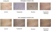 Fig. 3 The effect of nicotine and P. gingivalis on biomineralization on day 28. Groups 1 to 4 were grown in osteogenic media and groups 5 and 6 were grown in nonosteogenic media. (A, E) Group 1 (control), (B) group 2 (P. gingivalis lysate), (C) group 3 (nicotine), (D) group 4 (nicotine + P. gingivalis lysate), (F) group 5 (P. gingivalis alone), (G) group 6 (nicotine alone), and (H) nicotine + P. gingivalis.
Discussion
This study aims to study the differentiation of human PDL cells in response to nicotine and P. gingivalis. The conditions were selected to mimic the status of periodontal disease in smoking patients. MTT assay was used in this study since the assay has been well known to determine the cytotoxicity in cell culture system.[23] MTT cytotoxicity test showed that 50% inhibitory concentration of nicotine was 19.22 mM, which was similar to the 12.6 mM found in a previous study.[5] This study also found that P. gingivalis whole cell lysate at the concentration of 50 µg/mL was not toxic to PDLs similar to other studies.[24]
[25] Therefore, experiments were performed at 1 μM nicotine, a concentration similar to the plasma concentration, together with 50 µg/mL P. gingivalis lysate which showed no toxicity. The experiment were divided into eight groups: group 1 to group 4 were treated with odontogenic/osteogenic differentiation media, while group 5 to group 8 cells were cultured without odontogenic/osteogenic differentiation inducers. Group 1 acted as a negative control of cell differentiation, group 2 was to determine the effect of P. gingivalis lysate alone on cell differentiation, group 3 was to determine the effect of nicotine alone on cell differentiation, while group 4 was to determine the effect of both nicotine together with P. gingivalis lysate. Group 5 showed the normal growth of PDLs in the DMEM medium without osteogenic differentiation inducer. Group 6 showed the effect of P. gingivalis lysate on PDLs and group 7 showed the effect of nicotine alone on PDLs in DMEM. Finally, group 8 showed the effect of both P. gingivalis together with nicotine on PDLs in nondifferentiation medium. It was clearly showed that no mineralized nodule was found in nonosteogenic/odontogenic medium, while several nodules were found in osteogenic/odontogenic medium similar to other studies.[24]
Osteoinduction medium containing dexamethasone, ascorbic acid, and beta-glycerophosphate was used.[26] Gene expression was studied using four known osteogenic markers: Runx2, COL1A1, ALP, and OCN, comparing treatment and control groups. The results from groups 1 to 4 showed significant influence of P. gingivalis and nicotine on the osteogenic differentiation properties of PDL fibroblast, since the genes COL1A1, ALP, and Runx2 were decreased on day 7, while OCN gene expression was reduced on day 14.
Collagen, more specifically collagen type 1, is the most important and abundant organic component of the extracellular bone matrix that provides the bone strength and flexibility.[27] When the cell binds with collagen, mitogen-activated protein kinase is activated, which then transfers signaling to the nucleus, and activates transcription factor Runx2 by a phosphorylation process.[28]
ALP gene has been reported to be associated with osteogenic differentiation. Runx2 expression was found at the onset of differentiation, and regulated osteoblast-specific genes. It can facilitate the convergence of numerous osteogenic signaling pathways including OCN expression, which plays a key role in the differentiation of osteoblast progenitor cells.
The results from groups 1 to 4 also showed that in the presence of nicotine and/or P. gingivalis, osteogenic differentiation was inhibited, since the number and size of mineralized nodules on day 28 were reduced. The results were in good agreement with the gene expression data.
Nicotine exerts its cellular functions through the nicotinic acetylcholine receptors. The subunits of the nicotinic acetylcholine receptor are present in human MSC and PDL fibroblasts. In addition, nicotine altered the microRNA (miRNA) in nicotine-treated PDL stem cell which suggested that miRNAs might play an important role in the effects of nicotine on stem cell-associated healing delay in cigarette smoking.[29] A previous study reported that LPS from P. gingivalis could inhibit proliferation and differentiation of bone marrow stem cells.[9]
Porphyromonas gingivalis was also reported to affect cell proliferation and osteogenic differentiation of PDLs in vitro by downregulating Runx2 mRNA expression, and reduced mineral deposition. Other components of P. gingivalis such as lipid A also inhibited osteoblast function and gene expression through Toll-like receptor 2.[11]
Since P. gingivalis and nicotine have been implicated as a major etiologic factors in the development and progression of periodontitis. Periodontal pathogens and nicotine have been reported to induce oxidative stress via the endogenous superoxide mediation in PDLs.[5] This study is the first to clearly show that both nicotine and P. gingivalis together also reduced osteogenic differentiation of PDLs, which may occur in periodontal patients who regularly smoke, possibly leading to complications in healing of the periodontium and bone during treatment. This may help explain why treatment of regular smokers for chronic periodontal disease and periodontal regeneration are rather unpredictable. These results, together with results from other experiments, confirmed the negative effects of cigarette smoke on the PDL cells and their differentiation potential, especially in the presence of periodontal pathogens. Indeed, dentists should be aware of the problem of tissue regeneration in regular smokers, and consider more frequent follow-up. The mechanisms involved in the effect of bacteria and nicotine are still unclear, but could involve oxidative stress. Our results may be applied for the treatment of periodontal disease in smoking patients and may support the recommendation for periodontal patients to stop smoking.


