

Subscribe to RSS
DOI: 10.1055/s-0040-1713832
Endoscopic Ultrasound-Guided Transgastric Fine Needle Aspiration of Splenic Metastasis
Authors

Abstract
Splenic metastasis is extremely rare and is part of diffuse carcinomatosis. We present a case of a 60-year-old woman with transitional cell ovarian carcinoma who had hysterectomy with bilateral salpingo-oophorectomy in the past. She now presented with solitary splenic metastasis, confirmed by endoscopic ultrasound-guided transgastric fine needle aspiration biopsy.
Introduction
The word spleen comes from ancient Greek σπλήν (splén) and is not commonly affected by disease. The differential diagnosis of splenic masses include malignancy (lymphoma, metastatic disease), infection (tuberculosis, fungal), and infiltrative processes such as sarcoidosis.[1] Cross-sectional imaging can identify splenic masses, but they cannot differentiate the lesions. Histology is crucial but reserved only if there is no other safer accessible site to obtain tissue.[2] Splenic tissue samples can be obtained either by surgery (splenectomy), percutaneous biopsy, or endoscopic ultrasound (EUS) guidance.[3] The bleeding complication following percutaneous biopsy may be due to capsular laceration as a result of breathing.[4] Utilizing proximity to the gastrointestinal tract, EUS can sample a splenic mass, traversing less tissue, and may reduce the risk of hemorrhage.[5]
Metastasis in the spleen (breast, lung, ovarian, colorectal cancers and melanomas) occurs in approximately 1% of malignant tumors.[6] We present a rare case of a solitary splenic metastasis of transitional cell carcinoma of ovary, which occurred after a 4-year disease-free interval diagnosed by EUS-guided fine needle aspiration (FNA) biopsy.
Case Report
A 60-year-old woman presented with left upper quadrant pain for a duration of 5 days. In 2014, she was evaluated for right lower quadrant abdominal pain and fever for a duration of 30 days. She underwent staging laparotomy, total abdominal hysterectomy, and bilateral salpingo-oopherectomy for carcinoma ovary. She had received adjuvant chemotherapy with six cycles of paclitaxel and carboplatin. Follow-up imaging demonstrated complete remission of the disease. She had no other comorbidities or addictions. Her vital signs, physical examination, and laboratory results were unremarkable. Her blood cancer antigen 125 (CA125) level was 55.6 U/mL (reference range, 0–35 U/mL).
Abdominal contrast-enhanced CT was done, which revealed an ill-defined minimally enhancing hypodense lesion measuring 47 × 38 mm in size on the medial aspect of spleen ([Fig. 1]). After obtaining written informed consent, EUS was performed using a Pentax linear echoendoscope- EG-3870 UTK connected to a Hitachi Avius estiva ultrasound machine (2012). On EUS examination, there was a 3.5 × 3.3 cm heteroechoic mass lesion in the splenic hilum ([Fig. 2]). Transgastric EUS-guided FNA of the splenic lesion was done using a 22-G needle (EchoTip, Wilson-Cook, Winston-Salem, North Carolina, USA). The biopsy needle is placed under ultrasound guidance down to the splenic capsule. The patient then was instructed to hold breath in order to bring the lesion into the field of view and the biopsy trajectory. The needle is advanced into the lesion and four passes with thread biopsy were taken.[7] The needle was withdrawn and breathing resumed, decreasing the splenic capsular laceration/hematoma formation. Repeat EUS image showed contained needle tract hematoma ([Fig. 3]).


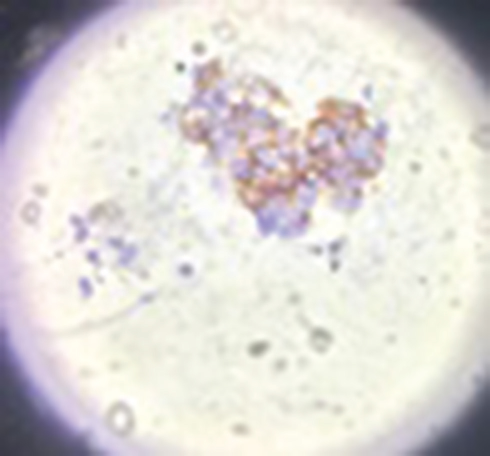
Histology revealed large atypical cells with hyperchromatic nuclei and moderate degree of pleomorphism ([Fig. 4]). Neoplastic cells showed positive immunohistochemical (ICH) staining for CK7 ([Fig. 5]) and CA 125 ([Fig. 6]) and were negative for CK 20 ([Fig. 7]) and Uroplakin III ([Fig. 8]). This immunophenotype is indicative of a metastatic primary transitional cell carcinoma of ovarian origin. She underwent uneventful open splenectomy, followed by adjuvant postoperative chemotherapy ([Figs. 9] [10]). She is asymptomatic at 1 month follow-up.


Discussion
Most splenic metastases are accompanied by multivisceral tumor dissemination. The rarity of splenic metastases might be explained by the following reasons: (1) the poorly developed lymphoid system of the spleen, especially the lack of afferent lymphatic vessels, prevents the spleen from receiving metastatic tumor cells via the lymphatic route; (2) the sharp angle of splenic artery branching from the celiac trunk inhibits large clumps of tumor cells from passing through; and (3) the microenvironment of the spleen may hinder the growth of micrometastatic foci (characteristically present after a period of clinical latency of 11–120 months).[6] [7] F-FDG positron emission tomography can distinguish benign from malignant masses of the spleen, but it is preferable to obtain a tissue diagnosis. As compared with surgery with the highest morbidity at 8.2%, image-guided percutaneous spleen biopsy has lower complication rate (4.2%). EUS-guided splenic biopsy is an emerging modality for tissue acquisition.[8]
Peripheral lesion away from hilum will be targeted in image-guided biopsy with some intervening normal splenic tissue to minimize bleeding risk[9]. EUS is better in inaccessible splenic lesions that are located closely to hilum.[10]
Isolated splenic metastasis like in our patient points to a favorable prognosis. The tumor showed positivity for CK7 and CA 125 which are typical of female genital tract. As the primary was transitional cell carcinoma of ovary, CK20 and Uroplakin III were done, which were negative, ruling out bladder metastasis.
In conclusion, EUS provides good images of the spleen through the gastric wall and can acquire tissue from inaccessible splenic lesions that are located closely to hilum. EUS-guided FNA has been reported to be useful modality for cytopathological diagnosis of splenic lesions.
Author Contributions
Avinash B. was involved in the concept of case report, manuscript preparation, and final approval.
Aravind S. Kapali was involved in the manuscript preparation and final approval.
Suneetha K.P. was involved in the manuscript preparation and final approval.
SatyaPrakash B.S. was involved in the manuscript preparation and final approval.
Declaration
No grants were received for this study.
Conflicts of Interest
The authors declare that there are no conflicts of interest regarding the publication of this paper.
-
References
- 1 McInnes MD, Kielar AZ, Macdonald DB. Percutaneous image-guided biopsy of the spleen: systematic review and meta-analysis of the complication rate and diagnostic accuracy. Radiology 2011; 260 (03) 699-708
- 2 Bastidas AB, Holloman D, Lankarani A, Nieto JM. Endoscopic ultrasound-guided Needle-Based Probe Confocal Laser Endomicroscopy (nCLE) of intrapancreatic ectopic spleen. ACG Case Rep J 2016; 3 (03) 196-198
- 3 Singh AK, Shankar S, Gervais DA, Hahn PF, Mueller PR. Image-guided percutaneous splenic interventions. Radiographics 2012; 32 (02) 523-534
- 4 Patel N, Dawe G, Tung K. Ultrasound-guided percutaneous splenic biopsy using an 18-G core biopsy needle: our experience with 52 cases. Br J Radiol 2015; 88 (1055) 20150400
- 5 Fritscher-Ravens A, Mylonaki M, Pantes A, Topalidis T, Thonke F, Swain P. Endoscopic ultrasound-guided biopsy for the diagnosis of focal lesions of the spleen. Am J Gastroenterol 2003; 98 (05) 1022-1027
- 6 Resta G, Vedana L, Marino S, Scagliarini L, Bandi M, Anania G. Isolated splenic metastasis of ovaric cancer. Case report and literature review. G Chir 2014; 35 (7-8) 181-184
- 7 Balekuduru AB, Dutta AK, Nagaruru SK, Sheik S, Kurella SP, Subbaraj SB. Comparison of diagnostic yield of endoscopic ultrasound-guided fine-needle aspiration cytology and cell block in solid lesions. J Dig Endosc 2017; 8: 176-181
- 8 Saab S, Challita Y, Holloman D, Hathaway K, Kahaleh M, Nieto J. Case series review of the safety and efficacy of endoscopic ultrasound-guided splenic mass core biopsy. Clin Endosc 2018; 51 (06) 600-601
- 9 Sammon J, Twomey M, Crush L, Maher MM, O’Connor OJ. Image-guided percutaneous splenic biopsy and drainage. Semin Intervent Radiol 2012; 29 (04) 301-310
- 10 Rana SS, Bhasin DK, Rao C. et al. Splenic tuberculosis diagnosed by endoscopic ultrasound-guided fine needle aspiration. Endosc Ultrasound 2012; 1 (03) 167-168
Address for correspondence
Publication History
Article published online:
22 June 2020
© .
Thieme Medical and Scientific Publishers Private Ltd.
A-12, Second Floor, Sector -2, NOIDA -201301, India
-
References
- 1 McInnes MD, Kielar AZ, Macdonald DB. Percutaneous image-guided biopsy of the spleen: systematic review and meta-analysis of the complication rate and diagnostic accuracy. Radiology 2011; 260 (03) 699-708
- 2 Bastidas AB, Holloman D, Lankarani A, Nieto JM. Endoscopic ultrasound-guided Needle-Based Probe Confocal Laser Endomicroscopy (nCLE) of intrapancreatic ectopic spleen. ACG Case Rep J 2016; 3 (03) 196-198
- 3 Singh AK, Shankar S, Gervais DA, Hahn PF, Mueller PR. Image-guided percutaneous splenic interventions. Radiographics 2012; 32 (02) 523-534
- 4 Patel N, Dawe G, Tung K. Ultrasound-guided percutaneous splenic biopsy using an 18-G core biopsy needle: our experience with 52 cases. Br J Radiol 2015; 88 (1055) 20150400
- 5 Fritscher-Ravens A, Mylonaki M, Pantes A, Topalidis T, Thonke F, Swain P. Endoscopic ultrasound-guided biopsy for the diagnosis of focal lesions of the spleen. Am J Gastroenterol 2003; 98 (05) 1022-1027
- 6 Resta G, Vedana L, Marino S, Scagliarini L, Bandi M, Anania G. Isolated splenic metastasis of ovaric cancer. Case report and literature review. G Chir 2014; 35 (7-8) 181-184
- 7 Balekuduru AB, Dutta AK, Nagaruru SK, Sheik S, Kurella SP, Subbaraj SB. Comparison of diagnostic yield of endoscopic ultrasound-guided fine-needle aspiration cytology and cell block in solid lesions. J Dig Endosc 2017; 8: 176-181
- 8 Saab S, Challita Y, Holloman D, Hathaway K, Kahaleh M, Nieto J. Case series review of the safety and efficacy of endoscopic ultrasound-guided splenic mass core biopsy. Clin Endosc 2018; 51 (06) 600-601
- 9 Sammon J, Twomey M, Crush L, Maher MM, O’Connor OJ. Image-guided percutaneous splenic biopsy and drainage. Semin Intervent Radiol 2012; 29 (04) 301-310
- 10 Rana SS, Bhasin DK, Rao C. et al. Splenic tuberculosis diagnosed by endoscopic ultrasound-guided fine needle aspiration. Endosc Ultrasound 2012; 1 (03) 167-168