Keywords
abducens nerve palsy - petroclival meningioma - intracranial aneurysm
Palavras-chave
paresia de nervo abducente - meningioma petroclival - aneurisma intracraniano
Introduction
After surgeries in places near the sixth abducens nerve, ipsilateral nerve palsy may
occur due to injury by surgical manipulation. However, palsy of the sixth cranial
nerve is also a rare complication following surgeries and procedures that can cause
cerebrospinal fluid (CSF) hypotension in areas distant from the nerve pathway. In
the literature, there are descriptions of this occurrence mainly in dural lesion spinal
surgeries[1]
[2] and dura mater puncture procedures,[3] such as lumbar punctures and spinal anesthesia.
The authors present a case of contralateral abducens nerve palsy after left middle
cerebral artery aneurysm surgery and left temporal meningioma, differing from most
of the literature reports describing lesions of the sixth cranial nerve following
procedures and surgeries of the spine. Our objective is to discuss the mechanisms
of the appearance of abducens nerve palsy when the site of manipulation is remote,
along with the particularities present in this case due to the presence of a petroclival
meningioma ipsilateral to the injured nerve.
Case Report
The patient is a 49-year-old woman. Her initial symptom was a headache. As part of
the diagnostic routine, a magnetic resonance imaging of the skull was requested. In
this examination, a left temporal meningioma, a left cerebral artery aneurysm and
a right petroclival meningioma were revealed ([Figs. 1], [2]).
 Fig. 1 Gadolinium-enhanced T1-weighted axial magnetic resonance image showing the left temporal
meningioma and the right petroclival meningioma.
Fig. 1 Gadolinium-enhanced T1-weighted axial magnetic resonance image showing the left temporal
meningioma and the right petroclival meningioma.
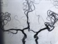 Fig. 2 Arteriography showing the left cerebral artery aneurysm.
After the identification of these lesions, the patient opted for the surgery of only
the left temporal meningioma and left cerebral artery aneurysm. For this, a pterional
craniotomy was performed on the left hemisphere of the brain.
In the postoperative period, the patient developed abducens nerve palsy on the right
side, contralateral to the surgery. After 3 months, the function of this nerve was
completely recovered spontaneously.
Discussion
Abducens nerve palsies in cases of procedures performed in distant sites were described
in the literature after lumbar puncture (diagnostic and therapeutic), inadvertent
lumbar puncture during epidural anesthesia, spinal anesthesia, CSF shunt, intrathecal
drug delivery, myelography, and lumbar drainage.[4] Cases of this occurrence have also been reported due to spinal surgery.[5] These procedures may lead to intracranial hypotension due to CSF drainage,[6] which would be the explanation for nerve damage. This is due to the anatomy and
the long course of the sixth cranial nerve and its relation to the base of the skull.
The abducens nerve innervates the lateral rectus muscle, whose function is the abduction
of the eye. Its nucleus lies on the dorsal surface of the pons ventrally to the floor
of the fourth ventricle. Throughout its course, the nerve has three points of pronounced
curvature.[7] When emerging from the lower part of the pons, the nerve performs its first sharp
curve in the upper direction. It then goes through the subarachnoid space superiorly
along the clivus and through the Dorello channel. This channel lies between the petrosphenoidal
ligament (Gruber ligament) and the apex of the petrous part of the temporal bone and
fixes the nerve.[8] Farther, the nerve curves for the second time, through the Dorello canal, around
the apex of the petrous part of the temporal bone and penetrates into the cavernous
sinus. In this region, it runs laterally to the internal carotid artery (around which
it makes its third curvature) and enters the orbit through the superior orbital fissure.
Soon after, it innervates the lateral rectus muscle.
In the course of its long intracranial path, the abducens nerve is subjected to mechanical
forces of traction and compression. In the aforementioned situations of intracranial
hypotension by CSF drainage, the nerve loses its support and remains stretched. This
is due to its anatomical relationships with the base of the skull and its long and
tortuous trajectory. The first sharp curve of the nerve in the petroclival region
when piercing the dura and entering the Dorello canal is the region most susceptible
to nerve stretching. In this area, the acute angle near the nerve fixation region
in the canal favors the risk of traction with the loss of brainstem support by intracranial
hypotension.[9] This stretching would injure the nerve, generating its palsy.
However, reports in the literature point to the palsy of the sixth cranial nerve by
this mechanism mostly through procedures and surgeries in the spine with dural puncture,
not intracranial surgeries. In this case, the drainage of the CSF and the opening
of the cisterns during surgery was sufficient to cause nerve damage.
We believe that the particularity of the case lies in the presence of the petroclival
meningioma ipsilateral to the injured nerve. The presence of petroclival meningioma
alone can generate an abducens nerve palsy due to traction and compression injury,[10] but in this case, the palsy manifested only after the aneurysm and temporal meningioma
surgery. As the nerve region susceptible to stretching is just along its tortuous
path through the petroclival region, the presence of the tumor in this area would
amplify the nerve traction. Thus, both the relative CSF imbalance and the consequent
loss of nerve support in the brainstem and the compression caused by the petroclival
meningioma were responsible, in this case, for the palsy of the nerve contralateral
to the surgery and ipsilateral to the unresected tumor.
Conclusion
Palsy of the abducens nerve, even when the site of manipulation is remote, is characterized
as a rare complication after surgeries and procedures with dural puncture. With relative
intracranial hypotension due to CSF imbalance, the nerve is subject to stretching
because of its long and tortuous course. Moreover, in this case, the petroclival meningioma
contralateral to the surgery may have contributed to transient abducens nerve palsy.


