

Subscribe to RSS
DOI: 10.1055/s-0033-1359120
Mega stents: a new option for management of leaks following laparoscopic sleeve gastrectomy
Authors
Corresponding author
Publication History
Publication Date:
12 February 2014 (online)

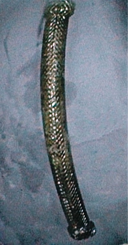
Postoperative leaks occur after laparoscopic sleeve gastrectomy (LSG) in 1 % – 3 % of cases [1]. Placement of covered metallic stents is an effective treatment strategy [2] [3], but in about 16.9 % of cases stent migration occurs [4]. To avoid the complication of stent migration, we have used a large stent (Niti-S Mega esophageal stent; Taewoong Medical, Gyeonggi-do, South Korea) in two patients who developed leaks following LSG. The first patient was a 50-year-old woman with a body mass index (BMI) of 35, who developed abdominal pain and vomiting 2 days after LSG. Contrast-enhanced computed tomography (CECT) of her abdomen revealed two perigastric collections with contrast leak ([Fig. 1]). Endoscopy showed a fistulous opening at the gastroesophageal junction ([Fig. 2]). A fully covered Mega esophageal stent (length 23 cm; diameter: body 24 mm, flanges 32 mm) was placed under fluoroscopic guidance with the proximal end in the esophagus and the distal end in suprapapillary position in the descending duodenum using a fluoroscopic marker, thus covering the entire stomach sleeve ([Fig. 3]). Repeat abdominal CECT revealed no collection at 8 weeks and the stent ([Fig. 4]) was removed.


The second patient was a 45-year-old woman (BMI 48.4) who developed abdominal pain, fever, and dyspnea on the 3rd postoperative day, requiring ventilatory support. Surgical re-exploration was done with augmentation of the staple line sutures. However, 15 days later abdominal CECT revealed a leak in the proximal part of the stomach. Endoscopy confirmed the site of the leak and this patient also underwent placement of a Mega esophageal stent. The stent was removed 6 weeks later after a contrast study showed no leak.
Conventional stents placed around gastroesophageal junction leaks would hang their lower end in the capacious antrum, enhancing the risk of migration. The Mega esophageal stent has the advantage of covering the entire stomach sleeve, which not only helps to cover the leak site but also decreases the risk of migration, since the lower end now rests in the duodenum. Even if the stent covers the ampulla of Vater, the bile and pancreatic juice can flow around the stent. Further studies in a larger number of patients would be helpful.
Endoscopy_UCTN_Code_TTT_1AR_2AZ
Competing interests: None
-
References
- 1 Clinical Issues Committee of the American Society for Metabolic and Bariatric surgery. Updated position statement on laparoscopic sleeve gastrectomy as a bariatric procedure. Surg Obes Relat Dis 2010; 6: 1-5
- 2 Casella G, Soricelli E, Rizzello M et al. Nonsurgical treatment of staple line leaks after laparoscopic sleeve gastrectomy. Obes Surg 2009; 19: 821-826
- 3 Serra C, Baltasar A, Andreo L et al. Treatment of gastric leaks with coated self-expanding stents after sleeve gastrectomy. Obes Surg 2007; 17: 866-872
- 4 Puli SR, Spofford IS, Thompson CC. Use of self-expandable stents in the treatment of bariatric surgery leaks: a systematic review and meta-analysis. Gastrointest Endosc 2012; 75: 287-293
Corresponding author
-
References
- 1 Clinical Issues Committee of the American Society for Metabolic and Bariatric surgery. Updated position statement on laparoscopic sleeve gastrectomy as a bariatric procedure. Surg Obes Relat Dis 2010; 6: 1-5
- 2 Casella G, Soricelli E, Rizzello M et al. Nonsurgical treatment of staple line leaks after laparoscopic sleeve gastrectomy. Obes Surg 2009; 19: 821-826
- 3 Serra C, Baltasar A, Andreo L et al. Treatment of gastric leaks with coated self-expanding stents after sleeve gastrectomy. Obes Surg 2007; 17: 866-872
- 4 Puli SR, Spofford IS, Thompson CC. Use of self-expandable stents in the treatment of bariatric surgery leaks: a systematic review and meta-analysis. Gastrointest Endosc 2012; 75: 287-293