

Subscribe to RSS
DOI: 10.1055/a-2164-0850
Color overlay of contrast-enhanced endoscopic ultrasound for pancreaticobiliary disease
Authors

Contrast-enhanced endoscopic ultrasound (CE-EUS) has been considered an important examination for visualization of blood flow and for its contribution to more accurate diagnosis in various conditions of the pancreaticobiliary region [1] [2] [3] [4] [5]. However, the conventional black and white mode may limit visual discernibility. A new EUS processor (EU-ME3; Olympus Co., Tokyo, Japan) has been equipped with a novel color overlay mode, which could potentially augment the perception of contrast agents, enhancing the utility of CE-EUS. We present three cases in which the color overlay mode of CE-EUS improved visualization during observation and tissue acquisition ([Video 1]).
Video 1 This video presents three cases in which the color overlay mode of contrast-enhanced endoscopic ultrasound improved visualization during observation and tissue acquisition.
Case 1: CE-EUS was performed for a patient with intraductal papillary mucinous neoplasm with nodules ([Fig. 1]). The color overlay mode offered a far clearer visualization of the contrast-enhanced nodules within the cyst, compared with the conventional mode ([Fig. 2], [Video 1]).


Case 2: A patient with intrahepatic cholangiocarcinoma whose lesion localization and boundaries were unclear with various imaging modalities underwent CE-EUS for observation. By utilizing color overlay mode, regions devoid of contrast agent within the tumor were better delineated, facilitating lesion localization ([Fig. 3]).

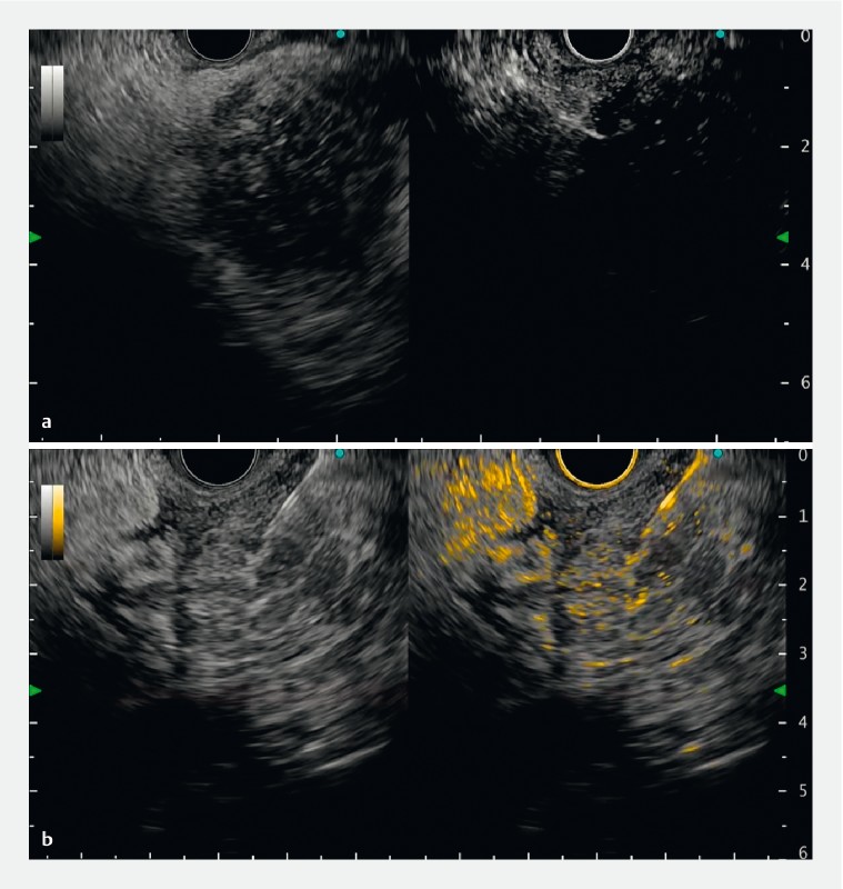
Case 3: A patient suspected of having an expansile necrotizing tumor in the head of the pancreas was scheduled for EUS-guided tissue acquisition ([Fig. 4]). Viable tissue sampling is paramount to enhance diagnostic yield. However, discernibility was challenging on conventional CE-EUS. By applying color overlay mode, contrast particles were clearly identified, leading to efficient sampling ([Fig. 5]).

Technological advances in endoscopic equipment allow endoscopists to perform the procedure more accurately. The newly introduced color overlay mode may increase accuracy and reduce endoscopists’ stress during EUS procedures.
Endoscopy_UCTN_Code_TTT_1AS
E-Videos is an open access online section of the journal Endoscopy, reporting on interesting cases and new techniques in gastroenterological endoscopy.
All papers include a high-quality video and are published with a Creative Commons
CC-BY license. Endoscopy E-Videos qualify for HINARI discounts and waivers and eligibility is automatically checked during the submission
process. We grant 100% waivers to articles whose corresponding authors are based in
Group A countries and 50% waivers to those who are based in Group B countries as classified
by Research4Life (see: https://www.research4life.org/access/eligibility/).
This section has its own submission website at https://mc.manuscriptcentral.com/e-videos
Competing interests
A. Katanuma has received honoraria as a lecture fee from Olympus Co., Tokyo, Japan. H. Toyonaga, T. Hayashi, M. Motoya, T. Kin, and K. Takahashi declare that they have no conflict of interest.
-
References
- 1 Gincul R, Palazzo M, Pujol B. et al. Contrast-harmonic endoscopic ultrasound for the diagnosis of pancreatic adenocarcinoma: a prospective multicenter trial. Endoscopy 2014; 46: 373-379
- 2 Kamata K, Kitano M, Omoto S. et al. Contrast-enhanced harmonic endoscopic ultrasonography for differential diagnosis of pancreatic cysts. Endoscopy 2016; 48: 35-41
- 3 Yamamoto N, Kato H, Tomoda T. et al. Contrast-enhanced harmonic endoscopic ultrasonography with time-intensity curve analysis for intraductal papillary mucinous neoplasms of the pancreas. Endoscopy 2016; 48: 26-34
- 4 Krishna SG, Rao BB, Ugbarugba E. et al. Diagnostic performance of endoscopic ultrasound for detection of pancreatic malignancy following an indeterminate multidetector CT scan: a systemic review and meta-analysis. Surg Endosc 2017; 31: 4558-4567
- 5 Yamashita Y, Shimokawa T, Ashida R. et al. Comparison of endoscopic ultrasonography with and without contrast enhancement for characterization of pancreatic tumors: a meta-analysis. Endosc Int Open 2022; 10: E369-E377
Corresponding author
Publication History
Article published online:
06 October 2023
© 2023. The Author(s). This is an open access article published by Thieme under the terms of the Creative Commons Attribution License, permitting unrestricted use, distribution, and reproduction so long as the original work is properly cited. (https://creativecommons.org/licenses/by/4.0/)
Georg Thieme Verlag KG
Rüdigerstraße 14, 70469 Stuttgart, Germany
-
References
- 1 Gincul R, Palazzo M, Pujol B. et al. Contrast-harmonic endoscopic ultrasound for the diagnosis of pancreatic adenocarcinoma: a prospective multicenter trial. Endoscopy 2014; 46: 373-379
- 2 Kamata K, Kitano M, Omoto S. et al. Contrast-enhanced harmonic endoscopic ultrasonography for differential diagnosis of pancreatic cysts. Endoscopy 2016; 48: 35-41
- 3 Yamamoto N, Kato H, Tomoda T. et al. Contrast-enhanced harmonic endoscopic ultrasonography with time-intensity curve analysis for intraductal papillary mucinous neoplasms of the pancreas. Endoscopy 2016; 48: 26-34
- 4 Krishna SG, Rao BB, Ugbarugba E. et al. Diagnostic performance of endoscopic ultrasound for detection of pancreatic malignancy following an indeterminate multidetector CT scan: a systemic review and meta-analysis. Surg Endosc 2017; 31: 4558-4567
- 5 Yamashita Y, Shimokawa T, Ashida R. et al. Comparison of endoscopic ultrasonography with and without contrast enhancement for characterization of pancreatic tumors: a meta-analysis. Endosc Int Open 2022; 10: E369-E377