

Subscribe to RSS
DOI: 10.1055/a-1922-6778
Head and Neck Ultrasound – EFSUMB Training Recommendations for the Practice of Medical Ultrasound in Europe
Authors


Abstract
Different surgical and medical specialists increasingly use head and neck ultrasound and ultrasound-guided interventions as part of their clinical practice. We need to ensure high quality and standardized practice across specialties, and this position paper of the European Federation of Societies for Ultrasound in Medicine and Biology (EFSUMB) describes the training requirements for head and neck ultrasound. Traditionally, a minimum number of ultrasound examinations indicates competence, but this is unreliable, and a general shift towards competence-based training is ongoing. For each EFSUMB level, we will outline the theoretical knowledge and skills needed for clinical practice. The recommendations follow the three EFSUMB competency levels for medical ultrasound practice. Level 1 describes the skills required to perform essential head and neck ultrasound examinations independently, level 2 includes ultrasound-guided interventions, while level 3 involves the practice of high-level neck ultrasound and use of advanced technologies. Our goal is to ensure high quality and standardized head and neck ultrasound practice performed by different clinical specialists with these recommendations.
Introduction to EFSUMB training levels
Ultrasound can provide detailed anatomical information of the head and neck and is the first-line imaging investigation for neck masses, lymph nodes, salivary gland, and thyroid nodules [1] [2]. Furthermore, ultrasound-guided tissue sampling is essential in the diagnostic workup of suspicious neck lesions [3] [4]. Advances in ultrasound technology have resulted in excellent image quality and have refined the opportunities for tissue characterization with multiparametric ultrasound [5]. The development of small portable ultrasound systems has resulted in increased availability for users outside radiology departments. Therefore, head and neck ultrasound is increasingly used by different surgical and medical specialists such as otorhinolaryngologists, endocrinologists, general surgeons, pediatricians, cytopathologists, emergency medicine physicians, nuclear medicine physicians, oncologists, and general practitioners [6] [7] [8] [9]. The increased use of ultrasound in the head and neck has the potential to improve patient care by allowing the physician responsible for treatment to use a diagnostic modality as an extension of the clinical examination [10] [11]. However, ultrasound is a complex skill, and an adequate degree of competency is required to carry out diagnostic ultrasound and ultrasound-guided interventions safely and effectively [12] [13] [] [15]. A systematic examination approach, good ultrasound technique, and focused reporting are preconditions for a high-quality head and neck ultrasound examination, and therefore should be included in the training [16] [16] [17] [19].
With the increased use of head and neck ultrasound, it is necessary to ensure an expected and reliable level of performance when being used by different specialties, and this position paper of the European Federation of Societies for Ultrasound in Medicine and Biology (EFSUMB) describes the training requirements for head and neck ultrasound in Europe. The recommendations will follow the general principles of three competency levels for the practice of medical ultrasound used by EFSUMB and the Royal College of Radiologists [20], [21]. In this position paper, we describe the three competency levels as:
-
Level 1 involves the knowledge and skills needed to perform essential head and neck ultrasound examinations independently.
-
Level 2 describes the skills needed to perform ultrasound-guided interventions. Therefore, practitioners who need to perform ultrasound-guided interventions in their clinical work would be expected to hold Level I and Level 2 competencies and skills.
-
Level 3 involves training and practice on a more advanced level and requires additional knowledge of advanced ultrasound technologies and engagement in education or research.
Traditionally, a minimum number of ultrasound examinations performed over time has been used to indicate competence. However, from research in medical education, we know that the number of procedures performed may not correlate to the competencies achieved as the learning curve for ultrasound is very variable for individuals [22]. A general shift towards competence-based education requirements accompanied by valid competency assessments has been emphasized and will be followed in these recommendations [23].
A good ultrasound scanning technique is a complex procedure, in which anatomical, clinical, and technical knowledge, proprioceptive skills, a systematic examination approach, accurate image interpretation, and documentation are needed to scan competently [15] [23] [24]. Training should, therefore, consist of theoretical teaching and practical modules [13] [26]. For each EFSUMB level, we will outline the theoretical knowledge and skills needed with a description of the corresponding training and assessment requirements for clinical practices.
Level 1
Knowledge base
Physics and instrumentation, ultrasound techniques and administration
-
The essential components of an ultrasound system
-
Types of transducers and their use
-
The production and description of the ultrasound image
-
Explanation of basic ultrasound controls (gain, depth, focus, frequency, etc.)
-
Knowledge of Doppler ultrasound (both color flow and power Doppler)
-
The recognition and explanation of common artifacts
-
Knowing the indications for head and neck ultrasound
-
Understanding the strengths, weaknesses, and limitations of head and neck ultrasound
-
Knowing the advantages/disadvantages and risks of ultrasound examination and intervention techniques in comparison to alternative diagnostic and interventional tools
For more details, please refer to the EFSUMB minimum training recommendations for medical ultrasound [21].
Knowledge of normal sonographic anatomy of the head and neck
Thyroid, major salivary glands, cervical lymph nodes (ultrasonographic architecture of lymph nodes and level classification), larynx and pharynx landmarks, major vessels, and the key muscular landmarks of the neck.
Knowledge of common pathologies of the head and neck
Thyroid
-
Characteristics of malignant thyroid nodules
-
Systematic nodule classification (e. g., EU-TIRADS/BTA U classification) and indications for when to perform an ultrasound-guided biopsy [27] [28].
Salivary gland
-
Differentiation between lymph nodes in the parotid gland and salivary gland neoplasia
-
Characteristics of benign and malignant salivary gland neoplasia
-
Characteristics of acute sialadenitis and sialolithiasis
Cervical lymph nodes
-
Characteristics of benign, reactive, and malignant lymph nodes (including B-mode, color, and power Doppler criteria)
-
Features of lymphadenopathy with abscess formation
-
Anatomical lymph node level classifications and patterns of lymphatic drainage [29] [30]
Competencies to be acquired
-
Ability to recognize important anatomical landmarks on head and neck ultrasound
-
Ability to perform a systematic ultrasound examination of the head and neck, including the thyroid, major salivary glands, and lymph node levels of the neck
-
Ability to perform ultrasound of neck structures and lesions on two planes
-
Ability to describe the location and characterization of neck lesions (echogenicity, margins, vascularization, calcification, shadowing, solid/cystic nature, etc.)
-
Ability to optimize an image (using gain, depth, focus, and frequency), to perform measurements, and to insert pictograms/annotations
-
Ability to evaluate thyroid nodules and to classify them accordingly using a systematic nodule classification system
-
Ability to differentiate between normal, reactive, and malignant lymph nodes (B-mode, color, and power Doppler)
-
Ability to perform color and power Doppler ultrasound and use it in head and neck imaging, especially for the characterization of lymph nodes
-
Ability to write a detailed report of the ultrasound findings, including grading, differential diagnoses, and conclusion where appropriate
-
Ability to perform comprehensive and standardized documentation of the ultrasound examination, including adequate acquisition and storage of images and video files
-
Recognize limitations of personal expertise and scanning conditions and know when to ask for more expert advice
Required training
To obtain level 1 certification, the trainee should demonstrate basic knowledge of the anatomy and pathologies of the head and neck in relation to ultrasound as well as practical skills demonstrating competency in performing a systematic ultrasound examination, image interpretation, documentation, and correct medical decision making based on the head and neck ultrasound examination [12] [24]. To ensure this knowledge and competence, we recommend:
-
First, trainees should attend formal head and neck ultrasound courses endorsed by EFSUMB or national ultrasound/medical societies. The core knowledge base described earlier could be taught through didactics, online material, or textbooks [31]. At least half of the course time should be reserved for “hands-on” ultrasound training on volunteers and real patient cases. Simulators or phantoms with typical pathological findings can partially substitute for real patient cases.
-
After participating in a head and neck ultrasound course, the trainee should perform a reasonable number of ultrasound examinations (depending on local/national requirements and practice under supervision) to qualify for a skills assessment for level 1 certification. The cases scanned should include an appropriate range of normal and abnormal cases, including palpable and impalpable lesions.
-
We recommend that mentorship and supervision of training should be provided by a practitioner who has reached at least EFSUMB level 2 competence or a comparable qualification of a national ultrasound or medical society.
Assessment of ultrasound competencies
The trainee should satisfactorily complete both knowledge and ultrasound skills assessment to ensure competency at EFSUMB level 1.
Knowledge assessment
A theoretical multiple-choice test should cover ultrasound physics, head and neck ultrasound anatomy, and head and neck ultrasound pathology as described earlier in the document. The basic knowledge for EFSUMB level 1 should preferably be passed before attending the course.
Practical skills assessment
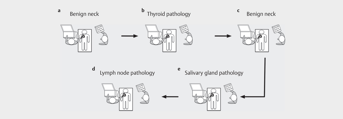
The competency assessment should include direct observation of the trainee performing a head and neck ultrasound examination. The ultrasound examination by the trainee should be assessed with a validated assessment tool, e. g., The Objective Structured Assessment of Ultrasound Skills (OSAUS) [12] [24], by a practitioner who has reached EFSUMB level 2 or corresponding national level. Observations of five systematic head and neck examinations of different head and neck ultrasound cases with a variation of benign and malignant head and neck findings are recommended to ensure reliable assessment [16], see [Fig. 1]. Video-recorded ultrasound exams could also be used as a more flexible alternative for the assessment of ultrasound skills if direct observation of performance is impossible [12] [32]. EFSUMB level 1 certification should be obtained when the trainee has followed the recommendations above and passed an EFSUMB-approved competency-based assessment.
Level 2
Level 2 focuses on the basic skills needed to safely perform ultrasound-guided fine-needle aspiration (FNA) biopsies, as it is an essential part of the diagnostic workup of most head and neck lesions. Depending on local/national practice and governance, it may be appropriate to include ultrasound-guided core biopsy in addition to ultrasound-guided FNA for level 2. Therefore, these skills may also be included during the training period for level 1, which focuses on diagnostic head and neck ultrasound skills.
Knowledge base
-
Ability to describe the principles of ultrasound-guided interventions and how to optimize needle visualization
-
Knowledge of the different techniques for ultrasound-guided interventions, including use of needle-guide or freehand technique (in-plane and out-of-plane technique)
-
Ability to handle the specimen depending on cytopathology/histopathology analyses needed, such as smearing on microscope slides, applying fixation techniques, and cell block or liquid-based cytology
-
Detailed knowledge of the principles of TNM staging of head and neck tumors and of other malignant tumors that commonly metastasize to cervical lymph nodes (esophageal and lung cancer, etc.)
Competencies to be acquired
Ability to perform safe and accurate ultrasound-guided fine-needle biopsies and/or ultrasound-guided core biopsies of head and neck lesions (thyroid nodules, lymph nodes, and salivary gland lesions).
Required training
Before performing invasive procedures on patients, initial training for ultrasound-guided interventions should be performed on phantoms to practice 3-dimensional hand-eye needle coordination.
Depending on local requirements and practice under supervision, a reasonable number of ultrasound-guided fine-needle aspirations (and/or core biopsies) should be performed based on EFSUMB guidelines [33]. It is preferable to do this with cytopathology support to provide feedback on the smear technique and to evaluate the final cytological/histological diagnosis. Finally, ultrasound-guided FNA or biopsy competence should be assessed with a validated assessment tool before level 2 certification is awarded [34].
Assessment of ultrasound competencies
Knowledge assessment
A theoretical multiple-choice test should cover basic principles for interventional ultrasound, techniques for collecting specimens for cytopathology/histopathology analyses, fixation techniques, and principles of TNM staging of head and neck cancer. The basic knowledge for EFSUMB level 2 should be assessed before performing a practical skills assessment.
Practical skills assessment
Direct observation of the trainee performing an ultrasound-guided procedure on a phantom should be carried out to ensure proper competency assessment. The ultrasound examination by the trainee should be assessed with an objective assessment tool by a practitioner who has reached at least EFSUMB level 2 or the corresponding national level [34]. If the trainee has followed the recommendations described above, the assessment of interventional ultrasound skills for EFSUMB level 2 can be performed simultaneously with the assessment of diagnostic ultrasound skills for EFSUMB level 1.
Level 3
Knowledge base
-
Knowledge of the principles of advanced ultrasound technologies such as elastography and contrast-enhanced ultrasound (CEUS) and their relevance for head and neck imaging
-
Knowledge of the anatomical, physiological, and developmental anomalies seen in the head and neck and how they present on ultrasound
-
Understanding of the principles behind selecting appropriate biopsy techniques in various head and neck conditions, e. g., the use of core biopsy in lymphoma
-
Ability to describe the ultrasound characteristics of rare pathologies of the head and neck (parathyroid adenomas, lymphatic malformation, carotid body paraganglioma, Schwannoma, vascular lesions, etc.)
-
Knowledge of the changes in ultrasound appearance associated with radiation and surgery
-
Knowledge of the latest developments and research in head and neck ultrasound
Competencies to be acquired
-
Ability to perform diagnostic head and neck ultrasound at an advanced level with a representative number of rare or demanding pathologies
-
Ability to use advanced technologies such as elastography or CEUS [35] [36]
-
Ability to perform a core biopsy in the head and neck
-
Ability to perform ultrasound examinations after radiotherapy and surgical procedures in the head and neck region and recognize therapy-induced ultrasound changes and differentiate them from residual/recurrent pathology
-
Acceptance of referrals from level 1 and level 2 practitioners and undertaking of more complex ultrasound examinations.
-
Mentoring and supervision of level 1 and 2 trainees and practitioners
-
Participation as an active ultrasound teacher or researcher
-
Ability to use required skills to develop advanced services under appropriate governance, e. g., ablation, injection, and marking techniques
Required training and assessment of competencies
Advanced diagnostic ultrasound skills are required, and physicians should regularly perform head and neck ultrasound to obtain level 3 certification. It is also a requirement to be involved in teaching and, if possible, ultrasound research. Candidate for level 3 certification need to document the above-described competencies, which should be assessed by EFSUMB experts in head and neck ultrasound.
Maintenance of skills
Practitioners should regularly perform or supervise head and neck examinations to maintain competence. There should be continuing professional development and updating of practice. Attendance at appropriate ultrasound courses or conferences of relevance to head and neck ultrasound, together with regular reviews of the current literature, is expected.
Conflict of Interest
Caroline Ewertsen, Christian Jenssen and Julian Kuenzel hold executive positions within EFSUMB.
Acknowledgements
We want to acknowledge Lynne Rudd for input and proofreading this manusscript.
-
References
- 1 McQueen AS, Bhatia KS. Head and neck ultrasound: technical advances, novel applications and the role of elastography. Clin Radiol 2018; 73: 81-93
- 2 Kunzel J, Strieth S, Wirth G, Bozzato A. Ultrasound in the Re-Staging of Cervical Metastases after Chemoradiotherapy for Head and Neck Cancer. Ultraschall Med 2018; 39: 659-666
- 3 Ahn D, Sohn JH, Yeo CK, Jeon JH. Feasibility of surgeon-performed ultrasound-guided core needle biopsy in the thyroid and lymph nodes. Head & neck 2016; 38: E1413-E1418
- 4 Todsen T, Bennedbaek FN, Kiss K, Hegedus L. Ultrasound-guided fine-needle aspiration biopsy of thyroid nodules. Head Neck 2021; 43: 1009-1013
- 5 Sidhu PS. Multiparametric Ultrasound (MPUS) Imaging: Terminology Describing the Many Aspects of Ultrasonography. Ultraschall Med 2015; 36: 315-317
- 6 Todsen T. Surgeon-performed ultrasonography. Dan Med J. 2017; 64
- 7 Hegedus L. Thyroid ultrasound. Endocrinology and metabolism clinics of North America 2001; 30: 339-360 viii-ix.
- 8 Jakowski JD. Pictorial Essay on How to Perform an Ultrasound-Guided Fine-Needle Aspiration Biopsy. AJSP: Reviews & Reports 2013; 18: 18
- 9 Lokkegaard T, Todsen T, Nayahangan LJ, Andersen CA, Jensen MB, Konge L. Point-of-care ultrasound for general practitioners: a systematic needs assessment. Scand J Prim Health Care 2020; 38: 3-11
- 10 Moore CL, Copel JA. Point-of-care ultrasonography. The New England journal of medicine 2011; 364: 749-757
- 11 Nielsen M, Cantisani V, Sidhu P, Badea R, Batko T, Carlsen J. et al. The Use of Handheld Ultrasound Devices – An EFSUMB Position Paper. Ultraschall in der Medizin - European Journal of Ultrasound. 2019; 40: 30-39
- 12 Todsen T, Melchiors J, Charabi B, Henriksen B, Ringsted C, Konge L. et al. Competency-based assessment in surgeon-performed head and neck ultrasonography: A validity study. The Laryngoscope 2018; 128: 1346-1352
- 13 Nicholls D, Sweet L, Hyett J. Psychomotor skills in medical ultrasound imaging: an analysis of the core skill set. Journal of ultrasound in medicine : official journal of the American Institute of Ultrasound in Medicine 2014; 33: 1349-1352
- 14 Lorentzen T, Nolsøe C, Ewertsen C, Nielsen M, Leen E, Havre R. et al. EFSUMB Guidelines on Interventional Ultrasound (INVUS), Part I. Ultraschall in der Medizin - European Journal of Ultrasound. 2015; 36: E3-E16
- 15 Wüstner M, Radzina M, Calliada F, Cantisani V, Havre RF, Jenderka KV. et al. Professional Standards in Medical Ultrasound - EFSUMB Position Paper (Long Version) – General Aspects. Ultraschall Med. 2022 Jul 18. doi: 10.1055/a-1857-4435. Online ahead of print.
- 16 Ernst BP, Katzer F, Kunzel J, Hodeib M, Strieth S, Eckrich J. et al. Impact of structured reporting on developing head and neck ultrasound skills. BMC Med Educ 2019; 19: 102
- 17 Ernst BP, Hodeib M, Strieth S, Kunzel J, Bischof F, Hackenberg B. et al. Structured reporting of head and neck ultrasound examinations. BMC Med Imaging 2019; 19: 25
- 18 Ernst BP, Strieth S, Katzer F, Hodeib M, Eckrich J, Bahr K. et al. The use of structured reporting of head and neck ultrasound ensures time-efficiency and report quality during residency. Eur Arch Otorhinolaryngol 2020; 277: 269-276
- 19 Trzebinska A, Dobruch-Sobczak K, Jakubowski W, Jedrzejowski M. Standards of the Polish Ultrasound Society – update. Ultrasound examination of thyroid gland and ultrasound-guided thyroid biopsy. J Ultrason 2014; 14: 49-60
- 20 The Royal College of Radiologists.. Ultrasound training recommendations for medical and surgical specialties. Third Edition. London, 2017
- 21 Education and Practical Standards Committee. European Federation of Societies for Ultrasound in Medicine and Biology. Minimum training recommendations for the practice of medical ultrasound. Ultraschall in der Medizin 2006; 27: 79-105
- 22 Jang TB, Ruggeri W, Dyne P, Kaji AH. The Learning Curve of Resident Physicians Using Emergency Ultrasonography for Cholelithiasis and Cholecystitis. Academic emergency medicine : official journal of the Society for Academic Emergency Medicine 2010; 17: 1247-1252
- 23 Holmboe ES, Sherbino J, Long DM, Swing SR, Frank JR. The role of assessment in competency-based medical education. Medical Teacher 2010; 32: 676-682
- 24 Tolsgaard MG, Todsen T, Sorensen JL, Ringsted C, Lorentzen T, Ottesen B. et al. International multispecialty consensus on how to evaluate ultrasound competence: a delphi consensus survey. PloS one 2013; 8: e57687
- 25 Kunzel J, Bozzato A, Ernst BP, Fuhrmann T, Ugele I, Scherl C. et al. [Quality in the appraisal of head and neck sonography results in university hospitals-a random sample]. HNO.. 2021
- 26 Todsen T, Jensen ML, Tolsgaard MG, Olsen BH, Henriksen BM, Hillingsø JG. et al. Transfer from point-of-care Ultrasonography training to diagnostic performance on patients-a randomized controlled trial. American journal of surgery 2016; 211: 40-45
- 27 Russ G, Bonnema SJ, Erdogan MF, Durante C, Ngu R, Leenhardt L. European Thyroid Association Guidelines for Ultrasound Malignancy Risk Stratification of Thyroid Nodules in Adults: The EU-TIRADS. European thyroid journal 2017; 6: 225-237
- 28 Weller A, Sharif B, Qarib MH, St Leger D, De Silva HS, Lingam RK. British Thyroid Association 2014 classification ultrasound scoring of thyroid nodules in predicting malignancy: Diagnostic performance and inter-observer agreement. Ultrasound. 2020; 28: 4-13
- 29 Som PM, Curtin HD, Mancuso AA. Imaging-based nodal classification for evaluation of neck metastatic adenopathy. AJR Am J Roentgenol 2000; 174: 837-844
- 30 van den Brekel MW, Castelijns JA. What the clinician wants to know: surgical perspective and ultrasound for lymph node imaging of the neck. Cancer Imaging 2005; 5 Spec No A S41-S49
- 31 Foss KT, Subhi Y, Aagaard R, Bessmann EL, Botker MT, Graumann O. et al. Developing an emergency ultrasound app - a collaborative project between clinicians from different universities. Scand J Trauma Resusc Emerg Med 2015; 23: 47
- 32 Subhi Y, Todsen T, Konge L. An integrable, web-based solution for easy assessment of video-recorded performances. Advances in medical education and practice 2014; 5: 103-105
- 33 Sidhu PS, Brabrand K, Cantisani V, Correas JM, Cui XW, D’Onofrio M. et al. EFSUMB Guidelines on Interventional Ultrasound (INVUS), Part II. Diagnostic Ultrasound-Guided Interventional Procedures (Short Version). Ultraschall Med 2015; 36: 566-580
- 34 Kahr Rasmussen N, Nayahangan LJ, Carlsen J, Ekberg O, Brabrand K, Albrecht-Beste E. et al. Evaluation of competence in ultrasound-guided procedures-a generic assessment tool developed through the Delphi method. Eur Radiol 2021; 31: 4203-4211
- 35 Sidhu P, Cantisani V, Dietrich C, Gilja O, Saftoiu A, Bartels E. et al. The EFSUMB Guidelines and Recommendations for the Clinical Practice of Contrast-Enhanced Ultrasound (CEUS) in Non-Hepatic Applications: Update 2017 (Long Version). Ultraschall in der Medizin - European Journal of Ultrasound 2018; 39: e2-e44
- 36 Saftoiu A, Gilja OH, Sidhu PS, Dietrich CF, Cantisani V, Amy D. et al. The EFSUMB Guidelines and Recommendations for the Clinical Practice of Elastography in Non-Hepatic Applications: Update 2018. Ultraschall Med 2019; 40: 425-453
Correspondence
Publication History
Received: 23 March 2022
Accepted: 27 July 2022
Article published online:
07 October 2022
© 2022. The Author(s). This is an open access article published by Thieme under the terms of the Creative Commons Attribution-NonDerivative-NonCommercial-License, permitting copying and reproduction so long as the original work is given appropriate credit. Contents may not be used for commercial purposes, or adapted, remixed, transformed or built upon. (https://creativecommons.org/licenses/by-nc-nd/4.0/).
Georg Thieme Verlag KG
Rüdigerstraße 14, 70469 Stuttgart, Germany
-
References
- 1 McQueen AS, Bhatia KS. Head and neck ultrasound: technical advances, novel applications and the role of elastography. Clin Radiol 2018; 73: 81-93
- 2 Kunzel J, Strieth S, Wirth G, Bozzato A. Ultrasound in the Re-Staging of Cervical Metastases after Chemoradiotherapy for Head and Neck Cancer. Ultraschall Med 2018; 39: 659-666
- 3 Ahn D, Sohn JH, Yeo CK, Jeon JH. Feasibility of surgeon-performed ultrasound-guided core needle biopsy in the thyroid and lymph nodes. Head & neck 2016; 38: E1413-E1418
- 4 Todsen T, Bennedbaek FN, Kiss K, Hegedus L. Ultrasound-guided fine-needle aspiration biopsy of thyroid nodules. Head Neck 2021; 43: 1009-1013
- 5 Sidhu PS. Multiparametric Ultrasound (MPUS) Imaging: Terminology Describing the Many Aspects of Ultrasonography. Ultraschall Med 2015; 36: 315-317
- 6 Todsen T. Surgeon-performed ultrasonography. Dan Med J. 2017; 64
- 7 Hegedus L. Thyroid ultrasound. Endocrinology and metabolism clinics of North America 2001; 30: 339-360 viii-ix.
- 8 Jakowski JD. Pictorial Essay on How to Perform an Ultrasound-Guided Fine-Needle Aspiration Biopsy. AJSP: Reviews & Reports 2013; 18: 18
- 9 Lokkegaard T, Todsen T, Nayahangan LJ, Andersen CA, Jensen MB, Konge L. Point-of-care ultrasound for general practitioners: a systematic needs assessment. Scand J Prim Health Care 2020; 38: 3-11
- 10 Moore CL, Copel JA. Point-of-care ultrasonography. The New England journal of medicine 2011; 364: 749-757
- 11 Nielsen M, Cantisani V, Sidhu P, Badea R, Batko T, Carlsen J. et al. The Use of Handheld Ultrasound Devices – An EFSUMB Position Paper. Ultraschall in der Medizin - European Journal of Ultrasound. 2019; 40: 30-39
- 12 Todsen T, Melchiors J, Charabi B, Henriksen B, Ringsted C, Konge L. et al. Competency-based assessment in surgeon-performed head and neck ultrasonography: A validity study. The Laryngoscope 2018; 128: 1346-1352
- 13 Nicholls D, Sweet L, Hyett J. Psychomotor skills in medical ultrasound imaging: an analysis of the core skill set. Journal of ultrasound in medicine : official journal of the American Institute of Ultrasound in Medicine 2014; 33: 1349-1352
- 14 Lorentzen T, Nolsøe C, Ewertsen C, Nielsen M, Leen E, Havre R. et al. EFSUMB Guidelines on Interventional Ultrasound (INVUS), Part I. Ultraschall in der Medizin - European Journal of Ultrasound. 2015; 36: E3-E16
- 15 Wüstner M, Radzina M, Calliada F, Cantisani V, Havre RF, Jenderka KV. et al. Professional Standards in Medical Ultrasound - EFSUMB Position Paper (Long Version) – General Aspects. Ultraschall Med. 2022 Jul 18. doi: 10.1055/a-1857-4435. Online ahead of print.
- 16 Ernst BP, Katzer F, Kunzel J, Hodeib M, Strieth S, Eckrich J. et al. Impact of structured reporting on developing head and neck ultrasound skills. BMC Med Educ 2019; 19: 102
- 17 Ernst BP, Hodeib M, Strieth S, Kunzel J, Bischof F, Hackenberg B. et al. Structured reporting of head and neck ultrasound examinations. BMC Med Imaging 2019; 19: 25
- 18 Ernst BP, Strieth S, Katzer F, Hodeib M, Eckrich J, Bahr K. et al. The use of structured reporting of head and neck ultrasound ensures time-efficiency and report quality during residency. Eur Arch Otorhinolaryngol 2020; 277: 269-276
- 19 Trzebinska A, Dobruch-Sobczak K, Jakubowski W, Jedrzejowski M. Standards of the Polish Ultrasound Society – update. Ultrasound examination of thyroid gland and ultrasound-guided thyroid biopsy. J Ultrason 2014; 14: 49-60
- 20 The Royal College of Radiologists.. Ultrasound training recommendations for medical and surgical specialties. Third Edition. London, 2017
- 21 Education and Practical Standards Committee. European Federation of Societies for Ultrasound in Medicine and Biology. Minimum training recommendations for the practice of medical ultrasound. Ultraschall in der Medizin 2006; 27: 79-105
- 22 Jang TB, Ruggeri W, Dyne P, Kaji AH. The Learning Curve of Resident Physicians Using Emergency Ultrasonography for Cholelithiasis and Cholecystitis. Academic emergency medicine : official journal of the Society for Academic Emergency Medicine 2010; 17: 1247-1252
- 23 Holmboe ES, Sherbino J, Long DM, Swing SR, Frank JR. The role of assessment in competency-based medical education. Medical Teacher 2010; 32: 676-682
- 24 Tolsgaard MG, Todsen T, Sorensen JL, Ringsted C, Lorentzen T, Ottesen B. et al. International multispecialty consensus on how to evaluate ultrasound competence: a delphi consensus survey. PloS one 2013; 8: e57687
- 25 Kunzel J, Bozzato A, Ernst BP, Fuhrmann T, Ugele I, Scherl C. et al. [Quality in the appraisal of head and neck sonography results in university hospitals-a random sample]. HNO.. 2021
- 26 Todsen T, Jensen ML, Tolsgaard MG, Olsen BH, Henriksen BM, Hillingsø JG. et al. Transfer from point-of-care Ultrasonography training to diagnostic performance on patients-a randomized controlled trial. American journal of surgery 2016; 211: 40-45
- 27 Russ G, Bonnema SJ, Erdogan MF, Durante C, Ngu R, Leenhardt L. European Thyroid Association Guidelines for Ultrasound Malignancy Risk Stratification of Thyroid Nodules in Adults: The EU-TIRADS. European thyroid journal 2017; 6: 225-237
- 28 Weller A, Sharif B, Qarib MH, St Leger D, De Silva HS, Lingam RK. British Thyroid Association 2014 classification ultrasound scoring of thyroid nodules in predicting malignancy: Diagnostic performance and inter-observer agreement. Ultrasound. 2020; 28: 4-13
- 29 Som PM, Curtin HD, Mancuso AA. Imaging-based nodal classification for evaluation of neck metastatic adenopathy. AJR Am J Roentgenol 2000; 174: 837-844
- 30 van den Brekel MW, Castelijns JA. What the clinician wants to know: surgical perspective and ultrasound for lymph node imaging of the neck. Cancer Imaging 2005; 5 Spec No A S41-S49
- 31 Foss KT, Subhi Y, Aagaard R, Bessmann EL, Botker MT, Graumann O. et al. Developing an emergency ultrasound app - a collaborative project between clinicians from different universities. Scand J Trauma Resusc Emerg Med 2015; 23: 47
- 32 Subhi Y, Todsen T, Konge L. An integrable, web-based solution for easy assessment of video-recorded performances. Advances in medical education and practice 2014; 5: 103-105
- 33 Sidhu PS, Brabrand K, Cantisani V, Correas JM, Cui XW, D’Onofrio M. et al. EFSUMB Guidelines on Interventional Ultrasound (INVUS), Part II. Diagnostic Ultrasound-Guided Interventional Procedures (Short Version). Ultraschall Med 2015; 36: 566-580
- 34 Kahr Rasmussen N, Nayahangan LJ, Carlsen J, Ekberg O, Brabrand K, Albrecht-Beste E. et al. Evaluation of competence in ultrasound-guided procedures-a generic assessment tool developed through the Delphi method. Eur Radiol 2021; 31: 4203-4211
- 35 Sidhu P, Cantisani V, Dietrich C, Gilja O, Saftoiu A, Bartels E. et al. The EFSUMB Guidelines and Recommendations for the Clinical Practice of Contrast-Enhanced Ultrasound (CEUS) in Non-Hepatic Applications: Update 2017 (Long Version). Ultraschall in der Medizin - European Journal of Ultrasound 2018; 39: e2-e44
- 36 Saftoiu A, Gilja OH, Sidhu PS, Dietrich CF, Cantisani V, Amy D. et al. The EFSUMB Guidelines and Recommendations for the Clinical Practice of Elastography in Non-Hepatic Applications: Update 2018. Ultraschall Med 2019; 40: 425-453