Subscribe to RSS
DOI: 10.1016/j.rboe.2017.10.010
Spondylectomy for Primary Ewing Lumbar Sarcoma in Children[*]
Article in several languages: português | English
Abstract
Primary Ewing sarcoma in the spine is very rare, and the treatment for it is multidisciplinary. There is no consensus regarding the optimal method of local control; however, en bloc resection is associated with an improvement in survival rates. The authors report a case of a 5-year-old girl who initially presented low back pain, and was diagnosed with Ewing sarcoma after being submitted to imaging studies by radiography, magnetic resonance and bone biopsy. A spondylectomy was performed in accordance with the Euro Ewing protocol. At the three-year follow-up, the patient had no restrictions regarding her daily activities, and there has been no evidence of recurrence to date.
#
Introduction
Ewing sarcoma was first described by James Ewing in 1921, and is the second most frequent malignant bone tumor in children, accounting for 3% of pediatric tumors and 10% of primary bone tumors.[1] The tumor typically occurs in the long bones (more often in the femur) and pelvis.[2] [3] [4] Primary involvement of the spine is rare, with a reported incidence of 5% of all primary sites.[2] [3]
The most common symptoms of Ewing sarcoma are localized pain and neurological impairment, ranging from radiculopathy to paraplegia.[1] [5] [6]
The imaging studies usually reveal aggressive vertebral body destruction, and most patients present with an extraosseous soft tissue mass with direct invasion of adjacent structures. Magnetic resonance imaging (MRI) is the method of choice for the detailed study of paravertebral involvement.[1] [5]
The surgical treatment is still challenging. En block resection is usually attempted, and is directly related to the prognosis.[2] [7]
Over the past three decades, the multidisciplinary approach and combined treatment with chemotherapy and radiotherapy have contributed to an improvement in the survival rates of the patients.[5] [7]
#
Case Report
A 5-year-old girl was observed in the Pediatric Emergency Service due to low back and abdominal pain, predominantly at night. Her symptoms had started one month prior to admission, with progressive back pain exacerbated in the previous two weeks. The pain did not respond to painkillers. The physical examination and the laboratory tests were unremarkable.
A radiograph of the lumbar spine was performed on admission, and it revealed a lytic lesion centered at L1 ([Fig. 1]). A subsequent MRI scan showed partial collapse of the L1 vertebral body due to an infiltrative lesion predominantly involving the right posterior half of the vertebral body and the ipsilateral pedicle, associated with a large extraosseous soft tissue mass with intracanal extension ([Fig. 2]). The imaging features were consistent with an aggressive process. The patient was submitted to a bone biopsy of L1, and, on the histological analysis, the morphological and immunophenotypic characteristics were consistent with Ewing sarcoma/primitive neuroectodermal tumor (PNET). A staging computed tomography (CT) scan did not reveal distant metastases.


After chemotherapy according to the Euro Ewing protocol, the patient underwent surgical treatment. A spondylectomy was performed in two stages under spinal cord neuromonitoring through somatosensory and motor-evoked potentials. In the first part of the procedure, we performed a pedicular fixation from D11-L3 and an excision of the posterior elements and pedicles of L1 via a posterior approach. In the second stage, the anterior corpectomy and reconstruction utilizing a Medtronic (Minneapolis, MN, US) cage filled with autologous iliac graft were performed via an anterior approach ([Fig. 3]).

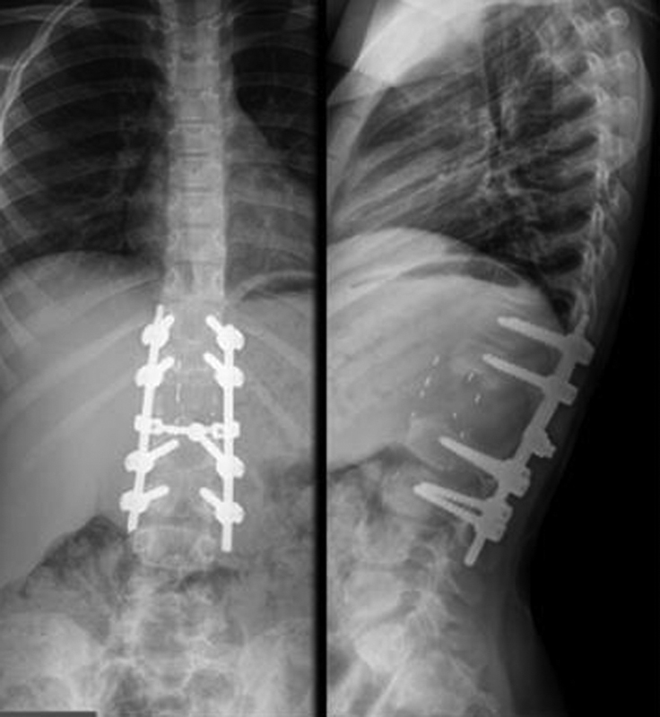
The patient was discharged 13 days postsurgery without neurological deficits. She continued the adjuvant treatment with chemotherapy and radiotherapy for 6 months, according to the protocol. On the first follow-up appointment, there was delayed healing of the surgical wound with necrosis of the edges, with need of extended dressing care for 4 weeks. There were no complications with the instrumentation ([Fig. 4]).

Over a three year period of follow-up, the patient had no restrictions regarding her daily activities, and there has been no evidence of recurrence to date. A follow-up CT scan revealed fusion of the instrumented levels ([Fig. 5]).

#
Discussion
The spine is often involved in cases of metastatic Ewing sarcoma. However, primary Ewing sarcomas in the spine are rare.[4] The imaging findings vary, but a destructive lytic lesion with partial collapse of the vertebral body is the most frequent presentation.[7] [8] Systemic manifestations such as fever and weight loss, accompanied by an increase in the total value of leucocytes and erythrocyte sedimentation, may be present.[1] [7] Given the variability of imaging features and variable phenotype, the differential diagnosis includes infection, neuroblastoma, eosinophilic granuloma and aneurysmal bone cyst.[7] The MRI plays a key role in the evaluation of tumor extension and in the preoperative planning.[1] [4] [8]
The treatment for Ewing Sarcoma is multidisciplinary. Most patients first diagnosed with a local disease actually present later with systemic micrometastases, which emphasizes the need for adjuvant treatment in all patients following local tumor surgical control.[1] Marco et al.[3] described a higher incidence of tumor recurrence and a lower survival rate in patients treated only with chemotherapy and radiotherapy compared with results reported by authors who performed the aggressive surgery. Therefore, aggressive surgical resection appears to improve the prognosis.[3]
The Tomita et al.[12] classification, which is based on the anatomical location of tumors in the axial and sagittal planes, is used as a guide for the surgical treatment. In the case described here, the tumor was classified as type 4, and the indicated surgical treatment was spondylectomy.[8]
There is still no consensus on the ideal method of local tumor control. Currently, en bloc resection with negative margins is accepted as the ideal treatment for these sarcomas, and is associated with an improvement in survival rates.[3] [7] [9] A systematic review published by Sciubba et al.[10] suggested that aggressive surgical resection was associated with improved overall survival rate and local control.[10]
The aim of en bloc vertebral resection is to enable complete tumor resection with free margins and decrease the likelihood of relapse.[5] [11] Tomita et al.[12] did not report any local recurrence in a total of 23 patients who underwent vertebral en bloc resection.[12]
Different techniques of en bloc resection have been described in the literature, such as the posterior technique or the combined posterior and anterior approaches.[11] However, en bloc resection is not always possible because of the proximity of the tumor to the neural structures.
In the current case, the authors preferred a two-stage approach. The posterior approach enabled the stabilization of two vertebral levels above and below L1 with pedicular screws, as well as the resection of the posterior elements of L1 with spinal decompression and release of the D12 and L1 nerve roots. In the second stage, via an anterior approach, the vertebral body and the remaining part of the pedicle were excised along with complete D12-L1 and L1-L2 discectomies, with better local control of the adjacent great vessels and viscera. This approach also enabled the reconstruction of the anterior column using an expandable cage. In a biomechanical study, Oda et al.[13] described that posterior pedicular instrumentation associated with anterior reconstruction with a cage provided the best stability after a spondylectomy.[13]
As a complication, our patient had necrosis of the surgical wound edges, which was previously described by other authors.[11] [14] Other frequent complications described in the literature include failure of the metallic hardware, pseudarthrosis or infection.[11]
In conclusion, primary Ewing sarcoma in the spine is rare. The treatment of these lesions remains a challenge, but tumor en bloc resection associated with an adjuvant treatment with radiotherapy and chemotherapy is associated with a better prognosis.
#
#
Conflict of Interests
The authors have no conflict of interests to declare.
* Study conducted at the Department of Orthopedic Surgery, Hospital de São João, Faculsade de Medicina, Universidade do Porto, Porto, Portugal. Originally Published by Elsevier.
-
Referências
- 1 Maheshwari AV, Cheng EY. Ewing sarcoma family of tumors. J Am Acad Orthop Surg 2010; 18 (02) 94-107
- 2 Sewell MD, Tan KA, Quraishi NA, Preda C, Varga PP, Williams R. Systematic review of en bloc resection in the management of Ewing's sarcoma of the mobile spine with respect to local control and disease-free survival. Medicine (Baltimore) 2015; 94 (27) e1019
- 3 Marco RA, Gentry JB, Rhines LD. et al. Ewing's sarcoma of the mobile spine. Spine 2005; 30 (07) 769-773
- 4 Feng H, Wang J, Guo P, Xu J, Feng J. Revision surgical treatment of a second lumbar Ewing Sarcoma: a report of a rare case. Medicine (Baltimore) 2015; 94 (30) e1190
- 5 Sciubba DM, Hsieh P, McLoughlin GS, Jallo GI. Pediatric tumors involving the spinal column. Neurosurg Clin N Am 2008; 19 (01) 81-92
- 6 Fenoy AJ, Greenlee JD, Menezes AH. et al. Primary bone tumors of the spine in children. J Neurosurg 2006; 105 (04) , Suppl): 252-260
- 7 Akeda K, Kasai Y, Kawakita E, Seto M, Kono T, Uchida A. Primary Ewing sarcoma of the spine mimicking a psoas abscess secondary to spinal infection. Spine 2009; 34 (09) E337-E341
- 8 Kim HJ, McLawhorn AS, Goldstein MJ, Boland PJ. Malignant osseous tumors of the pediatric spine. J Am Acad Orthop Surg 2012; 20 (10) 646-656
- 9 Rao G, Suki D, Chakrabarti I. et al. Surgical management of primary and metastatic sarcoma of the mobile spine. J Neurosurg Spine 2008; 9 (02) 120-128
- 10 Sciubba DM, Okuno SH, Dekutoski MB, Gokaslan ZL. Ewing and osteogenic sarcoma: evidence for multidisciplinary management. Spine 2009; 34 (22) , Suppl): S58-S68
- 11 Liljenqvist U, Lerner T, Halm H, Buerger H, Gosheger G, Winkelmann W. En bloc spondylectomy in malignant tumors of the spine. Eur Spine J 2008; 17 (04) 600-609
- 12 Tomita K, Kawahara N, Baba H, Tsuchiya H, Fujita T, Toribatake Y. Total en bloc spondylectomy. A new surgical technique for primary malignant vertebral tumors. Spine 1997; 22 (03) 324-333
- 13 Oda I, Cunningham BW, Abumi K, Kaneda K, McAfee PC. The stability of reconstruction methods after thoracolumbar total spondylectomy. An in vitro investigation. Spine 1999; 24 (16) 1634-1638
- 14 Krepler P, Windhager R, Bretschneider W, Toma CD, Kotz R. Total vertebrectomy for primary malignant tumours of the spine. J Bone Joint Surg Br 2002; 84 (05) 712-715
Address for correspondence
Publication History
Received: 06 September 2017
Accepted: 19 October 2017
Article published online:
03 February 2020
© 2020. The Author(s). This is an open access article published by Thieme under the terms of the Creative Commons Attribution-NonDerivative-NonCommercial-License, permitting copying and reproduction so long as the original work is given appropriate credit. Contents may not be used for commercial purposes, or adapted, remixed, transformed or built upon. (https://creativecommons.org/licenses/by-nc-nd/4.0/).
Sociedade Brasileira de Ortopedia e Traumatologia. Published by Thieme Revinter Publicações Ltda
Rio de Janeiro, Brazil
-
Referências
- 1 Maheshwari AV, Cheng EY. Ewing sarcoma family of tumors. J Am Acad Orthop Surg 2010; 18 (02) 94-107
- 2 Sewell MD, Tan KA, Quraishi NA, Preda C, Varga PP, Williams R. Systematic review of en bloc resection in the management of Ewing's sarcoma of the mobile spine with respect to local control and disease-free survival. Medicine (Baltimore) 2015; 94 (27) e1019
- 3 Marco RA, Gentry JB, Rhines LD. et al. Ewing's sarcoma of the mobile spine. Spine 2005; 30 (07) 769-773
- 4 Feng H, Wang J, Guo P, Xu J, Feng J. Revision surgical treatment of a second lumbar Ewing Sarcoma: a report of a rare case. Medicine (Baltimore) 2015; 94 (30) e1190
- 5 Sciubba DM, Hsieh P, McLoughlin GS, Jallo GI. Pediatric tumors involving the spinal column. Neurosurg Clin N Am 2008; 19 (01) 81-92
- 6 Fenoy AJ, Greenlee JD, Menezes AH. et al. Primary bone tumors of the spine in children. J Neurosurg 2006; 105 (04) , Suppl): 252-260
- 7 Akeda K, Kasai Y, Kawakita E, Seto M, Kono T, Uchida A. Primary Ewing sarcoma of the spine mimicking a psoas abscess secondary to spinal infection. Spine 2009; 34 (09) E337-E341
- 8 Kim HJ, McLawhorn AS, Goldstein MJ, Boland PJ. Malignant osseous tumors of the pediatric spine. J Am Acad Orthop Surg 2012; 20 (10) 646-656
- 9 Rao G, Suki D, Chakrabarti I. et al. Surgical management of primary and metastatic sarcoma of the mobile spine. J Neurosurg Spine 2008; 9 (02) 120-128
- 10 Sciubba DM, Okuno SH, Dekutoski MB, Gokaslan ZL. Ewing and osteogenic sarcoma: evidence for multidisciplinary management. Spine 2009; 34 (22) , Suppl): S58-S68
- 11 Liljenqvist U, Lerner T, Halm H, Buerger H, Gosheger G, Winkelmann W. En bloc spondylectomy in malignant tumors of the spine. Eur Spine J 2008; 17 (04) 600-609
- 12 Tomita K, Kawahara N, Baba H, Tsuchiya H, Fujita T, Toribatake Y. Total en bloc spondylectomy. A new surgical technique for primary malignant vertebral tumors. Spine 1997; 22 (03) 324-333
- 13 Oda I, Cunningham BW, Abumi K, Kaneda K, McAfee PC. The stability of reconstruction methods after thoracolumbar total spondylectomy. An in vitro investigation. Spine 1999; 24 (16) 1634-1638
- 14 Krepler P, Windhager R, Bretschneider W, Toma CD, Kotz R. Total vertebrectomy for primary malignant tumours of the spine. J Bone Joint Surg Br 2002; 84 (05) 712-715