
RSS-Feed abonnieren
Bitte kopieren Sie die angezeigte URL und fügen sie dann in Ihren RSS-Reader ein.
https://www.thieme-connect.de/rss/thieme/de/10.1055-s-00000118.xml
Radiologie up2date 2015; 15(01): 9-12
DOI: 10.1055/s-0034-1391277
DOI: 10.1055/s-0034-1391277
Der besondere Fall
Unklare Leberherde bei Sigmadivertikulitis
Weitere Informationen
Publikationsverlauf
Publikationsdatum:
10. März 2015 (online)

Fallvorstellung
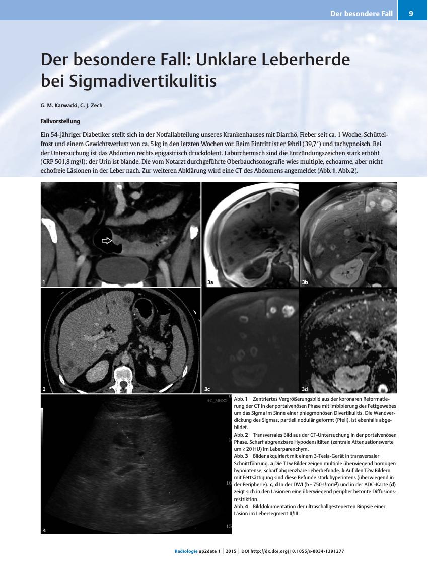
Ein 54-jähriger Diabetiker stellt sich in der Notfallabteilung unseres Krankenhauses mit Diarrhö, Fieber seit ca. 1 Woche, Schüttelfrost und einem Gewichtsverlust von ca. 5 kg in den letzten Wochen vor. Beim Eintritt ist er febril (39,7°) und tachypnoisch. Bei der Untersuchung ist das Abdomen rechts epigastrisch druckdolent. Laborchemisch sind die Entzündungszeichen stark erhöht (CRP 501,8 mg/l); der Urin ist blande. Die vom Notarzt durchgeführte Oberbauchsonografie wies multiple, echoarme, aber nicht echofreie Läsionen in der Leber nach. Zur weiteren Abklärung wird eine CT des Abdomens angemeldet (Abb. [1], Abb. [2]).
-
Literatur
- 1 Stollman N, Raskin JB. Diverticular disease of the colon. Lancet 2004; 363: 631-639
- 2 Chapman J, Davies M, Wolff B et al. Complicated Diverticulitis. Ann Surg 2005; 242: 576-583
- 3 Riemann JF, Fischbach W, Galle PR. Leberabszess. In: Gastroenterologie in Klinik und Praxis: Das komplette Referenzwerk für Klinik und Praxis. Stuttgart: Thieme; 2007: 1336-1339
- 4 Krige JEJ, Beckingham IJ. Liver abscesses and hydatid disease. BMJ 2001; 322: 537-540
- 5 Murarka S, Pranav F, Dandavate V. Pyogenic Liver Abscess Secondary to Disseminated Streptococcus Anginosus from Sigmoid Diverticulitis. J Glob Infect Dis 2011; 3: 79-81
- 6 Balci NC, Semelka RC, Noone TC et al. Pyogenic hepatic abscesses: MRI findings on T1- and T2-weighted and serial gadolinium-enhanced gradient-echo images. J Magn Reson Imaging JMRI 1999; 9: 285-290
- 7 Namasivayam S, Martin DR, Saini S. Imaging of liver metastases: MRI. Cancer Imaging 2007; 7: 2-9
- 8 Chan JHM, Tsui EYK, Luk SH et al. Diffusion-weighted MR imaging of the liver: distinguishing hepatic abscess from cystic or necrotic tumor. Abdom Imaging 2001; 26: 161-165
- 9 Park HJ, Kim SH, Jang KM et al. Differentiating hepatic abscess from malignant mimickers: Value of diffusion-weighted imaging with an emphasis on the periphery of the lesion: Hepatic Abscess Versus Malignant Mimickers. J Magn Reson Imaging 2013; 38: 1333-1341
- 10 Holzapfel K, Rummeny E, Gaa J. Diffusion-weighted MR imaging of hepatic abscesses: possibility of different apparent diffusion coefficient (ADC)-values in early and mature abscess formation. Abdom Imaging 2007; 32: 538-539