

Subscribe to RSS
DOI: 10.1055/a-2740-3836
Parenchymal loop technique using a novel 0.018-inch guidewire in endoscopic ultrasound-guided pancreatic duct drainage
Authors

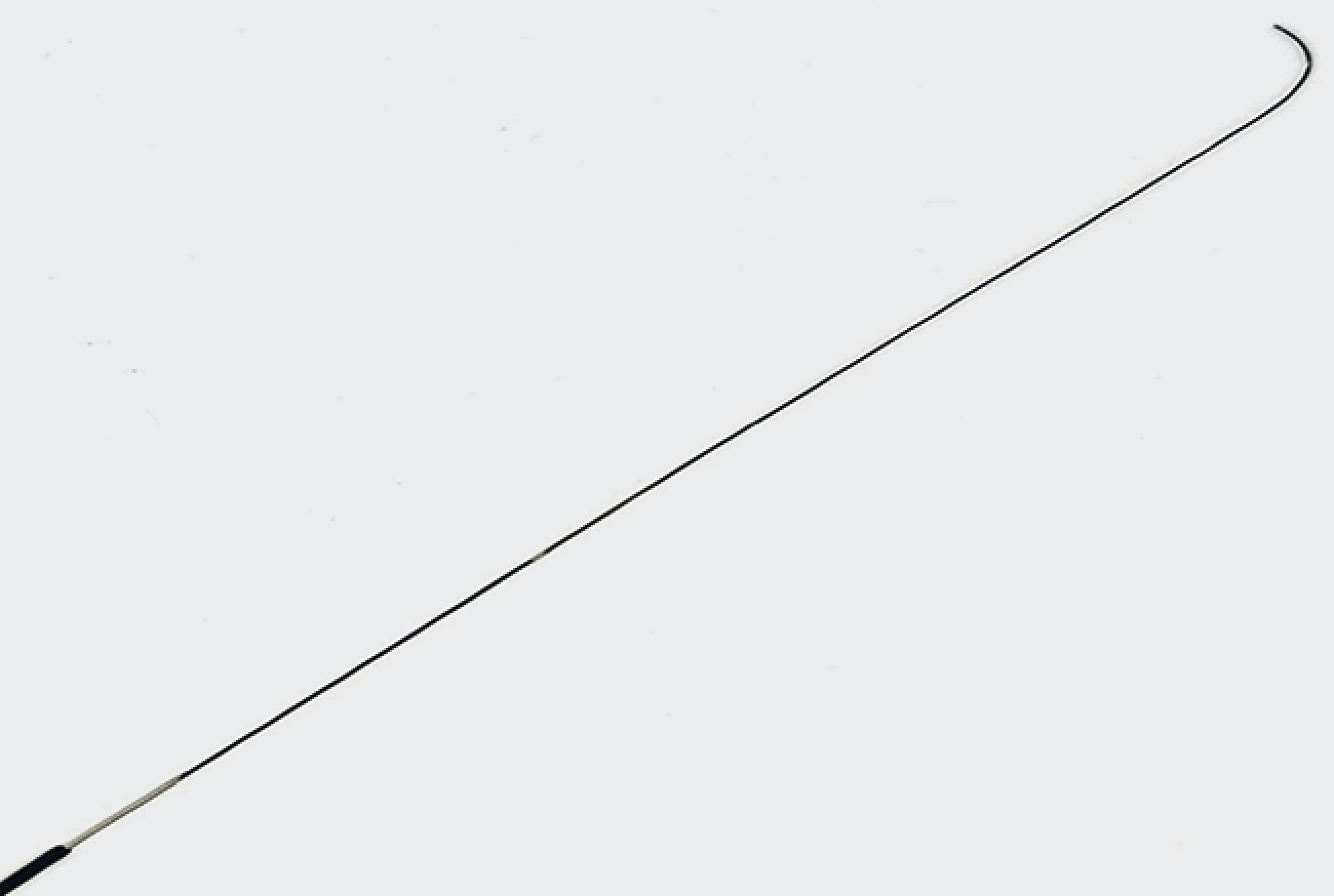
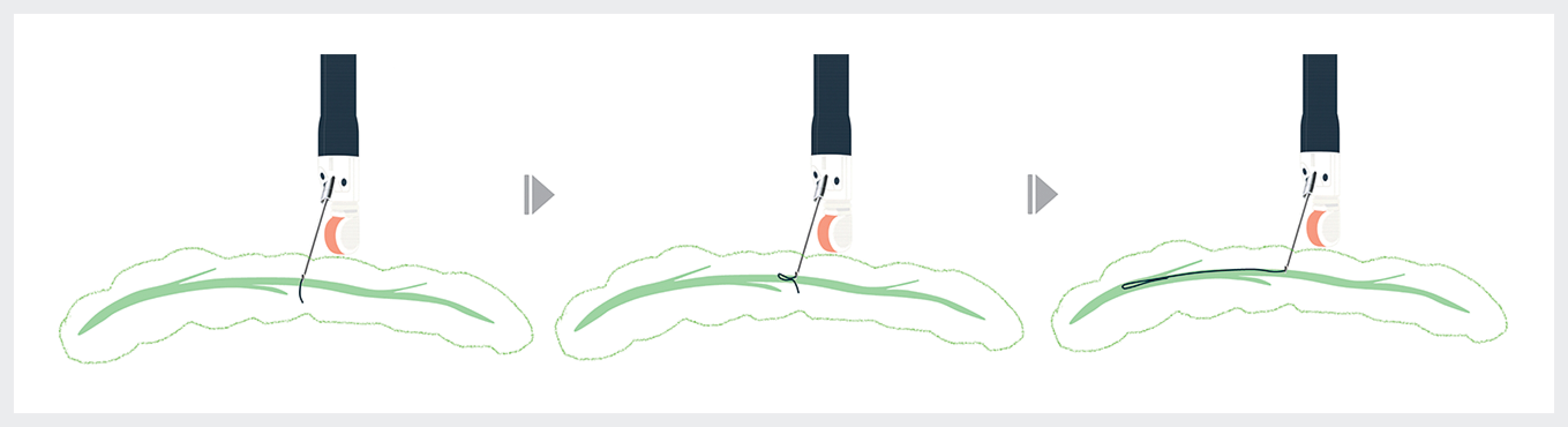
Endoscopic ultrasound-guided pancreatic duct drainage (EUS-PDD) is indicated when transpapillary pancreatic duct drainage is unsuccessful [1] [2]. EUS-PDD with a 22-gauge needle is effective for non-dilated pancreatic ducts; however, it is limited to a 0.018-inch guidewire. Because of its small caliber, a 0.018-inch guidewire easily migrates outside of the pancreatic duct through the puncture tract, making the procedure difficult and causing guidewire shearing [3] [4]. A novel 0.018-inch guidewire (J-wire premier NM; J-Mit, Kyoto, Japan) features a highly flexible and extended tip, which facilitates loop formation. In addition, the absence of visible markers reduces resistance within the needle, allowing safe withdrawal ([Fig. 1]). These characteristics enable what we term the “parenchymal loop technique”. In this approach, the guidewire, even after migration into the parenchyma, can be advanced into the pancreatic duct by forming a loop ([Fig. 2] and [Video 1]).
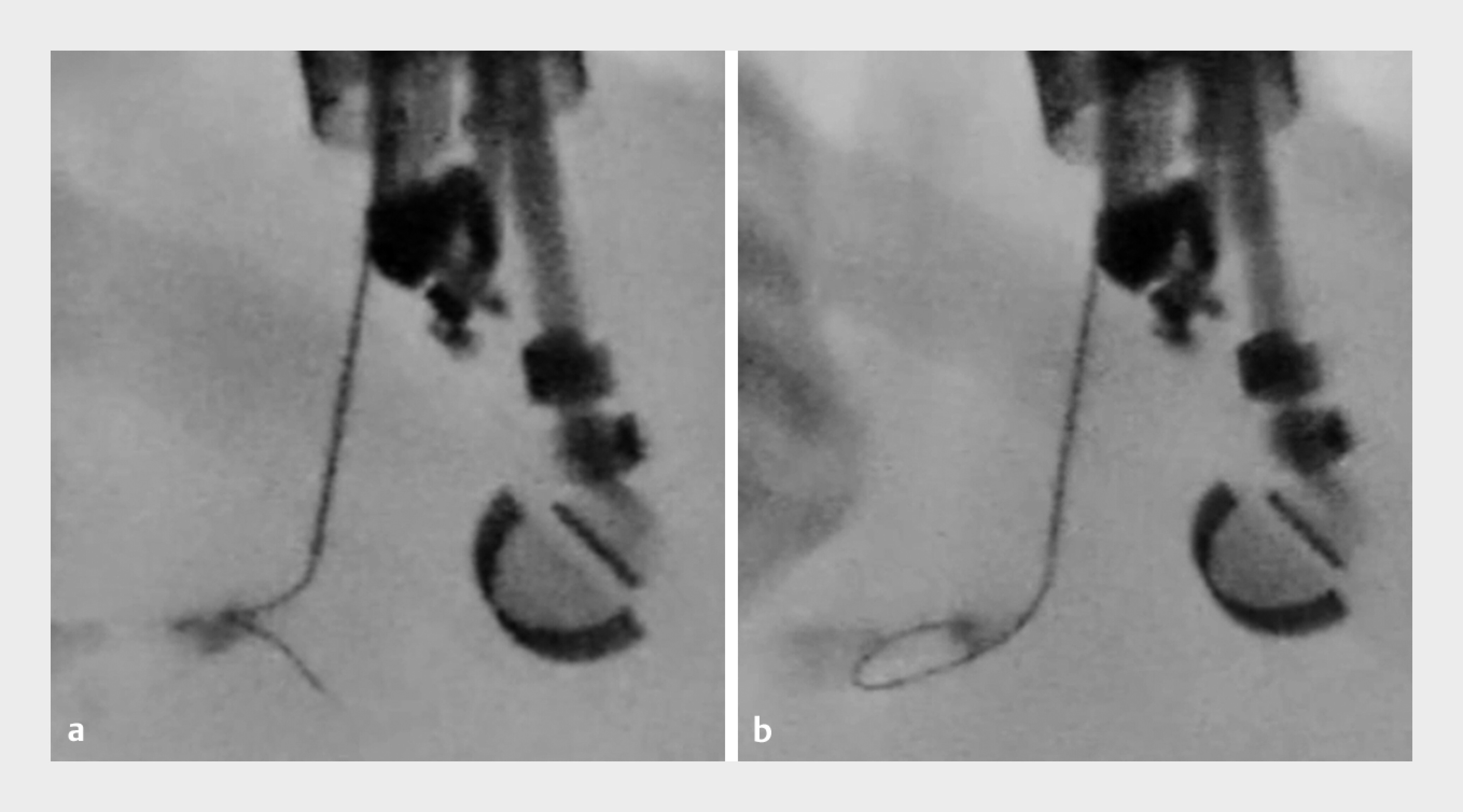
A 63-year-old man with chronic pancreatitis was admitted to our hospital with symptomatic pancreatic duct stones. Because the transpapillary approach was difficult owing to duodenal edema, EUS-PDD was performed ([Fig. 3]). First, the main pancreatic duct, measuring only 1.1 mm on EUS, was punctured with a 22-gauge needle. After puncture, the contrast medium was injected, and the novel 0.018-inch guidewire was introduced. Although the guidewire repeatedly migrated into pancreatic parenchyma, it could be withdrawn without stacking. When the tip migrated into the parenchyma, gentle further advancement caused it to be pushed back and inverted, leading to loop formation and enabling successful insertion into the main pancreatic duct ([Fig. 4]). The tract was dilated with a drill dilator, and the 0.018-inch guidewire was exchanged for a 0.025-inch guidewire. Finally, a 7-Fr plastic stent (Through&Pass Type IT; Gadelius Medical, Tokyo, Japan) was successfully deployed.

To the best of our knowledge, this is the first report of EUS-PDD achieved by the parenchymal loop technique using a novel 0.018-inch guidewire.
Endoscopy_UCTN_Code_TTT_1AS_2AD
E-Videos is an open access online section of the journal Endoscopy, reporting on interesting cases and new techniques in gastroenterological endoscopy.
All papers include a high-quality video and are published with a Creative Commons
CC-BY license. Endoscopy E-Videos qualify for HINARI discounts and waivers and eligibility is automatically checked during the submission
process. We grant 100% waivers to articles whose corresponding authors are based in
Group A countries and 50% waivers to those who are based in Group B countries as classified
by Research4Life (see: https://www.research4life.org/access/eligibility/).
This section has its own submission website at https://mc.manuscriptcentral.com/e-videos.
Publication History
Article published online:
28 November 2025
© 2025. The Author(s). This is an open access article published by Thieme under the terms of the Creative Commons Attribution License, permitting unrestricted use, distribution, and reproduction so long as the original work is properly cited. (https://creativecommons.org/licenses/by/4.0/).
Georg Thieme Verlag KG
Oswald-Hesse-Straße 50, 70469 Stuttgart, Germany
-
References
- 1 Imoto A, Ogura T, Higuchi K. et al. Endoscopic ultrasound-guided pancreatic duct drainage: techniques and literature review of transmural stenting. Clin Endosc 2020; 53: 525-534
- 2 Devière J. Endoscopic ultrasound-guided pancreatic duct interventions. Gastrointest Endosc Clin N Am 2023; 33: 845-854
- 3 Ogura T, Ueno S, Okuda A. et al. Expanding indications for endoscopic ultrasound-guided hepaticogastrostomy for patients with insufficient dilatation of the intrahepatic bile duct using a 22G needle combined with a novel 0.018-inch guidewire (with video). Digest Endosc 2021; 33: 1126-1133
- 4 Vila JJ, Pérez-Miranda M, Vazquez-Sequeiros E. et al. Initial experience with EUS-guided cholangiopancreatography for biliary and pancreatic duct drainage: a Spanish national survey. Gastrointest Endosc 2012; 76: 1133-1141